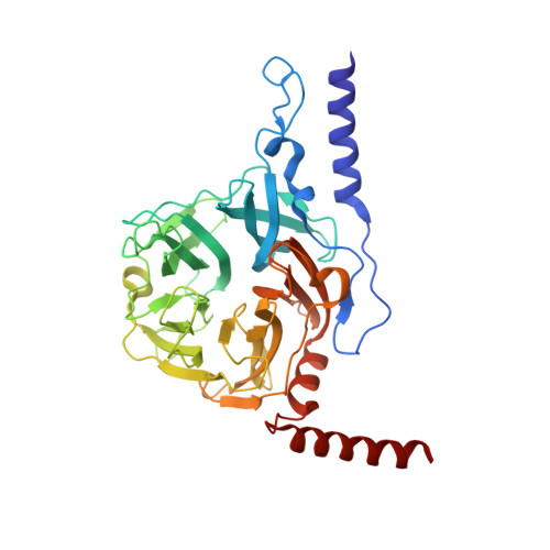
3WAT

Crystal structure of 4-O-beta-D-mannosyl-D-glucose phosphorylase MGP complexed with Man+Glc


Experimental Data Snapshot

  • Method: X-RAY DIFFRACTION
  • Resolution: 1.60 Å
  • R-Value Free: 0.175 
  • R-Value Work: 0.162 
  • R-Value Observed: 0.163 

Starting Model: experimental
View more details

wwPDB Validation   3D Report Full Report


Ligand Structure Quality Assessment 


This is version 1.3 of the entry. See complete history


Literature

Structure of Novel Enzyme in Mannan Biodegradation Process 4-O-beta-d-Mannosyl-d-Glucose Phosphorylase MGP

Nakae, S.Ito, S.Higa, M.Senoura, T.Wasaki, J.Hijikata, A.Shionyu, M.Ito, S.Shirai, T.

(2013) J Mol Biol 425: 4468-4478

  • DOI: https://doi.org/10.1016/j.jmb.2013.08.002
  • Primary Citation of Related Structures:  
    3WAS, 3WAT, 3WAU, 4KMI

  • PubMed Abstract: 

    The crystal structure of a novel component of the mannan biodegradation system, 4-O-β-D-mannosyl-D-glucose phosphorylase (MGP), was determined to a 1.68-Å resolution. The structure of the enzyme revealed a unique homohexameric structure, which was formed by using two helices attached to the N-terminus and C-terminus as a tab for sticking between subunits. The structures of MGP complexes with genuine substrates, 4-O-β-D-mannosyl-D-glucose and phosphate, and the product D-mannose-1-phosphate were also determined. The complex structures revealed that the invariant residue Asp131, which is supposed to be the general acid/base, did not exist close to the glycosidic Glc-O4 atom, which should be protonated in the catalytic reaction. Also, no solvent molecule that might mediate a proton transfer from Asp131 was observed in the substrate complex structure, suggesting that the catalytic mechanism of MGP is different from those of known disaccharide phosphorylases.


  • Organizational Affiliation

    Department of Bioscience, Nagahama Institute of Bio-science and Technology, and Japan Science and Technology Agency Institute for Bioinformatics Research and Development, 1266 Tamura, Nagahama 526-0829, Japan.


Macromolecules
Find similar proteins by:  (by identity cutoff)  |  3D Structure
Entity ID: 1
MoleculeChains Sequence LengthOrganismDetailsImage
4-O-beta-D-mannosyl-D-glucose phosphorylase
A, B
390Bacteroides fragilis NCTC 9343Mutation(s): 0 
Gene Names: BF0772
EC: 2.4.1.281
UniProt
Find proteins for Q5LH68 (Bacteroides fragilis (strain ATCC 25285 / DSM 2151 / CCUG 4856 / JCM 11019 / LMG 10263 / NCTC 9343 / Onslow / VPI 2553 / EN-2))
Explore Q5LH68 
Go to UniProtKB:  Q5LH68
Entity Groups  
Sequence Clusters30% Identity50% Identity70% Identity90% Identity95% Identity100% Identity
UniProt GroupQ5LH68
Sequence Annotations
Expand
  • Reference Sequence
Small Molecules
Ligands 3 Unique
IDChains Name / Formula / InChI Key2D Diagram3D Interactions
BGC
Query on BGC

Download Ideal Coordinates CCD File 
E [auth A],
I [auth B]
beta-D-glucopyranose
C6 H12 O6
WQZGKKKJIJFFOK-VFUOTHLCSA-N
BMA
Query on BMA

Download Ideal Coordinates CCD File 
F [auth A],
J [auth B]
beta-D-mannopyranose
C6 H12 O6
WQZGKKKJIJFFOK-RWOPYEJCSA-N
PO4
Query on PO4

Download Ideal Coordinates CCD File 
C [auth A],
D [auth A],
G [auth B],
H [auth B]
PHOSPHATE ION
O4 P
NBIIXXVUZAFLBC-UHFFFAOYSA-K
Experimental Data & Validation

Experimental Data

  • Method: X-RAY DIFFRACTION
  • Resolution: 1.60 Å
  • R-Value Free: 0.175 
  • R-Value Work: 0.162 
  • R-Value Observed: 0.163 
  • Space Group: H 3
Unit Cell:
Length ( Å )Angle ( ˚ )
a = 82.54α = 90
b = 82.54β = 90
c = 307.48γ = 120
Software Package:
Software NamePurpose
HKL-2000data collection
SOLVEphasing
PHENIXrefinement
MOSFLMdata reduction
SCALEPACKdata scaling

Structure Validation

View Full Validation Report


Ligand Structure Quality Assessment 


Entry History 

Deposition Data

Revision History  (Full details and data files)

  • Version 1.0: 2013-09-04
    Type: Initial release
  • Version 1.1: 2013-11-20
    Changes: Database references
  • Version 1.2: 2020-07-29
    Type: Remediation
    Reason: Carbohydrate remediation
    Changes: Data collection, Derived calculations, Structure summary
  • Version 1.3: 2023-11-08
    Changes: Data collection, Database references, Refinement description, Structure summary