3V11

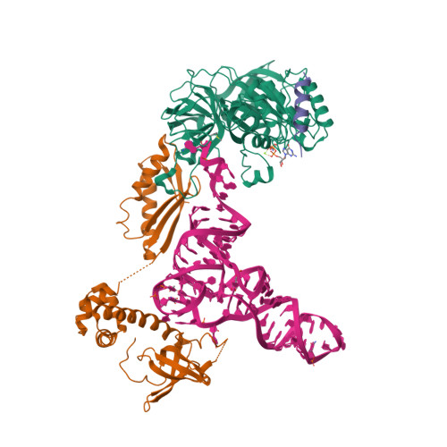
Structure of the ternary initiation complex AIF2:GDPNP:methionylated initiator TRNA


Experimental Data Snapshot

  • Method: X-RAY DIFFRACTION
  • Resolution: 5.00 Å
  • R-Value Free: 0.344 
  • R-Value Work: 0.262 
  • R-Value Observed: 0.265 

Starting Models: experimental
View more details

wwPDB Validation   3D Report Full Report


This is version 1.3 of the entry. See complete history


Literature

Structure of the ternary initiation complex aIF2-GDPNP-methionylated initiator tRNA.

Schmitt, E.Panvert, M.Lazennec-Schurdevin, C.Coureux, P.D.Perez, J.Thompson, A.Mechulam, Y.

(2012) Nat Struct Mol Biol 19: 450-454

  • DOI: https://doi.org/10.1038/nsmb.2259
  • Primary Citation of Related Structures:  
    3V11

  • PubMed Abstract: 

    Eukaryotic and archaeal translation initiation factor 2 (e/aIF2) is a heterotrimeric GTPase that has a crucial role in the selection of the correct start codon on messenger RNA. We report the 5-Å resolution crystal structure of the ternary complex formed by archaeal aIF2 from Sulfolobus solfataricus, the GTP analog GDPNP and methionylated initiator tRNA. The 3D model is further supported by solution studies using small-angle X-ray scattering. The tRNA is bound by the α and γ subunits of aIF2. Contacts involve the elbow of the tRNA and the minor groove of the acceptor stem, but not the T-stem minor groove. We conclude that despite considerable structural homology between the core γ subunit of aIF2 and the elongation factor EF1A, these two G proteins of the translation apparatus use very different tRNA-binding strategies.


  • Organizational Affiliation

    Laboratoire de Biochimie, Unité mixte de Recherche 7654, Ecole Polytechnique, Centre National de la Recherche Scientifique, Palaiseau, France. emma@botrytis.polytechnique.fr


Macromolecules

Find similar proteins by:  (by identity cutoff)  |  3D Structure
Entity ID: 1
MoleculeChains Sequence LengthOrganismDetailsImage
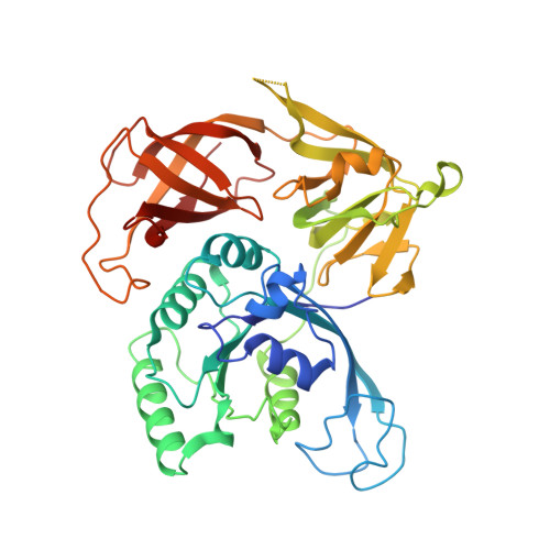
Translation initiation factor 2 subunit gamma414Saccharolobus solfataricus P2Mutation(s): 0 
Gene Names: eif2gSSO0412
EC: 3.6.5.3
UniProt
Find proteins for Q980A5 (Saccharolobus solfataricus (strain ATCC 35092 / DSM 1617 / JCM 11322 / P2))
Explore Q980A5 
Go to UniProtKB:  Q980A5
Entity Groups  
Sequence Clusters30% Identity50% Identity70% Identity90% Identity95% Identity100% Identity
UniProt GroupQ980A5
Sequence Annotations
Expand
  • Reference Sequence
Find similar proteins by:  (by identity cutoff)  |  3D Structure
Entity ID: 2
MoleculeChains Sequence LengthOrganismDetailsImage
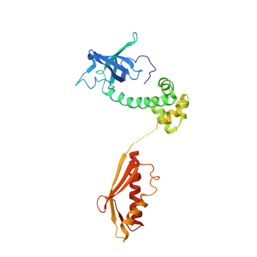
Translation initiation factor 2 subunit alpha266Saccharolobus solfataricus P2Mutation(s): 0 
Gene Names: eif2aSSO1050
UniProt
Find proteins for Q97Z79 (Saccharolobus solfataricus (strain ATCC 35092 / DSM 1617 / JCM 11322 / P2))
Explore Q97Z79 
Go to UniProtKB:  Q97Z79
Entity Groups  
Sequence Clusters30% Identity50% Identity70% Identity90% Identity95% Identity100% Identity
UniProt GroupQ97Z79
Sequence Annotations
Expand
  • Reference Sequence
Find similar proteins by:  (by identity cutoff)  |  3D Structure
Entity ID: 3
MoleculeChains Sequence LengthOrganismDetailsImage
Translation initiation factor 2 subunit beta138Saccharolobus solfataricus P2Mutation(s): 0 
Gene Names: eif2bSSO2381
UniProt
Find proteins for Q97W59 (Saccharolobus solfataricus (strain ATCC 35092 / DSM 1617 / JCM 11322 / P2))
Explore Q97W59 
Go to UniProtKB:  Q97W59
Entity Groups  
Sequence Clusters30% Identity50% Identity70% Identity90% Identity95% Identity100% Identity
UniProt GroupQ97W59
Sequence Annotations
Expand
  • Reference Sequence
Find similar nucleic acids by:  (by identity cutoff)  |  3D Structure
Entity ID: 4
MoleculeChains LengthOrganismImage
Initiator tRNA77Escherichia coli
Sequence Annotations
Expand
  • Reference Sequence
Small Molecules
Ligands 3 Unique
IDChains Name / Formula / InChI Key2D Diagram3D Interactions
GNP
Query on GNP

Download Ideal Coordinates CCD File 
F [auth A]PHOSPHOAMINOPHOSPHONIC ACID-GUANYLATE ESTER
C10 H17 N6 O13 P3
UQABYHGXWYXDTK-UUOKFMHZSA-N
MET
Query on MET

Download Ideal Coordinates CCD File 
G [auth D]METHIONINE
C5 H11 N O2 S
FFEARJCKVFRZRR-BYPYZUCNSA-N
MG
Query on MG

Download Ideal Coordinates CCD File 
E [auth A]MAGNESIUM ION
Mg
JLVVSXFLKOJNIY-UHFFFAOYSA-N
Experimental Data & Validation

Experimental Data

  • Method: X-RAY DIFFRACTION
  • Resolution: 5.00 Å
  • R-Value Free: 0.344 
  • R-Value Work: 0.262 
  • R-Value Observed: 0.265 
  • Space Group: P 32 2 1
Unit Cell:
Length ( Å )Angle ( ˚ )
a = 133α = 90
b = 133β = 90
c = 167.2γ = 120
Software Package:
Software NamePurpose
PHASERphasing
PHENIXrefinement
XDSdata reduction
XSCALEdata scaling

Structure Validation

View Full Validation Report


Entry History 

Deposition Data

Revision History  (Full details and data files)

  • Version 1.0: 2012-03-28
    Type: Initial release
  • Version 1.1: 2012-04-18
    Changes: Database references
  • Version 1.2: 2017-11-08
    Changes: Refinement description
  • Version 1.3: 2023-09-13
    Changes: Data collection, Database references, Derived calculations, Refinement description