3U2P

Crystal structure of N-terminal three extracellular domains of ErbB4/Her4


Experimental Data Snapshot

  • Method: X-RAY DIFFRACTION
  • Resolution: 2.57 Å
  • R-Value Free: 0.243 
  • R-Value Work: 0.192 
  • R-Value Observed: 0.195 

Starting Model: experimental
View more details

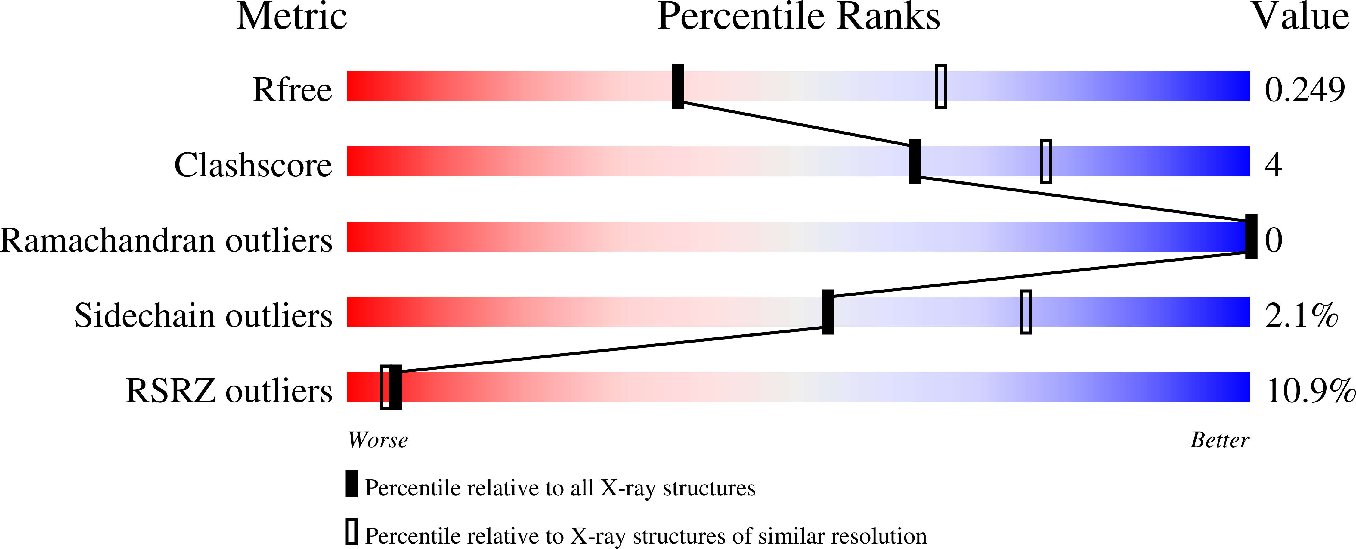
wwPDB Validation   3D Report Full Report


Ligand Structure Quality Assessment 


This is version 1.3 of the entry. See complete history


Literature

The ErbB4 extracellular region retains a tethered-like conformation in the absence of the tether.

Liu, P.Bouyain, S.Eigenbrot, C.Leahy, D.J.

(2012) Protein Sci 21: 152-155

  • DOI: https://doi.org/10.1002/pro.753
  • Primary Citation of Related Structures:  
    3U2P

  • PubMed Abstract: 

    The epidermal growth factor receptor (EGFR) and its homologs ErbB3 and ErbB4 adopt a tethered conformation in the absence of ligand in which an extended hairpin loop from domain II contacts the juxtamembrane region of domain IV and tethers the domain I/II pair to the domain III/IV pair. By burying the hairpin loop, which is required for formation of active receptor dimers, the tether contact was thought to prevent constitutive activation of EGFR and its homologs. Amino-acid substitutions at key sites within the tether contact region fail to result in constitutively active receptors however. We report here the 2.5 Å crystal structure of the N-terminal three extracellular domains of ErbB4, which bind ligand but lack domain IV and thus the tether contact. This ErbB4 fragment nonetheless adopts a domain arrangement very similar to the arrangement adopted in the presence of the tether suggesting that regions in addition to the tether contribute to maintaining this conformation and inactivity in the absence of the tether contact. We suggest that the tether conformation may have evolved to prevent crosstalk between different EGFR homologs and thus allow diversification of EGFR and its homologs.


  • Organizational Affiliation

    Department of Biophysics and Biophysical Chemistry, Johns Hopkins University School of Medicine, 725 N. Wolfe St., Baltimore, MD 21205, USA.


Macromolecules
Find similar proteins by:  (by identity cutoff)  |  3D Structure
Entity ID: 1
MoleculeChains Sequence LengthOrganismDetailsImage
Receptor tyrosine-protein kinase erbB-4497Homo sapiensMutation(s): 0 
Gene Names: ERBB4HER4
EC: 2.7.10.1
UniProt & NIH Common Fund Data Resources
Find proteins for Q15303 (Homo sapiens)
Explore Q15303 
Go to UniProtKB:  Q15303
PHAROS:  Q15303
GTEx:  ENSG00000178568 
Entity Groups  
Sequence Clusters30% Identity50% Identity70% Identity90% Identity95% Identity100% Identity
UniProt GroupQ15303
Glycosylation
Glycosylation Sites: 5Go to GlyGen: Q15303-1
Sequence Annotations
Expand
  • Reference Sequence
Experimental Data & Validation

Experimental Data

  • Method: X-RAY DIFFRACTION
  • Resolution: 2.57 Å
  • R-Value Free: 0.243 
  • R-Value Work: 0.192 
  • R-Value Observed: 0.195 
  • Space Group: P 21 21 21
Unit Cell:
Length ( Å )Angle ( ˚ )
a = 42.17α = 90
b = 85.56β = 90
c = 152.4γ = 90
Software Package:
Software NamePurpose
HKL-2000data collection
PHASERphasing
BUSTERrefinement
HKL-2000data reduction
HKL-2000data scaling

Structure Validation

View Full Validation Report


Ligand Structure Quality Assessment 


Entry History 

Deposition Data

Revision History  (Full details and data files)

  • Version 1.0: 2011-11-09
    Type: Initial release
  • Version 1.1: 2012-10-03
    Changes: Database references
  • Version 1.2: 2020-07-29
    Type: Remediation
    Reason: Carbohydrate remediation
    Changes: Data collection, Derived calculations, Structure summary
  • Version 1.3: 2023-09-13
    Changes: Data collection, Database references, Refinement description, Structure summary