3TS5

Crystal Structure of a Light Chain Domain of Scallop Smooth Muscle Myosin


Experimental Data Snapshot

  • Method: X-RAY DIFFRACTION
  • Resolution: 2.39 Å
  • R-Value Free: 0.241 
  • R-Value Work: 0.191 
  • R-Value Observed: 0.195 

wwPDB Validation   3D Report Full Report


This is version 1.1 of the entry. See complete history


Literature

Crystal structure of a phosphorylated light chain domain of scallop smooth-muscle Myosin.

Senthil Kumar, V.S.O'Neall-Hennessey, E.Reshetnikova, L.Brown, J.H.Robinson, H.Szent-Gyorgyi, A.G.Cohen, C.

(2011) Biophys J 101: 2185-2189

  • DOI: https://doi.org/10.1016/j.bpj.2011.09.028
  • Primary Citation of Related Structures:  
    3TS5, 3TUY

  • PubMed Abstract: 

    We have determined the crystal structure of a phosphorylated smooth-muscle myosin light chain domain (LCD). This reconstituted LCD is of a sea scallop catch muscle myosin with its phosphorylatable regulatory light chain (RLC SmoA). In the crystal structure, Arg(16), an arginine residue that is present in this isoform but not in vertebrate smooth-muscle RLC, stabilizes the phosphorylation site. This arginine interacts with the carbonyl group of the phosphorylation-site serine in the unphosphorylated LCD (determined previously), and with the phosphate group when the serine is phosphorylated. However, the overall conformation of the LCD is essentially unchanged upon phosphorylation. This result provides additional evidence that phosphorylation of the RLC is unlikely to act as an on-switch in regulation of scallop catch muscle myosin.


  • Organizational Affiliation

    Rosenstiel Basic Medical Sciences Research Center, Brandeis University, Waltham, Massachusetts, USA.


Macromolecules
Find similar proteins by:  (by identity cutoff)  |  3D Structure
Entity ID: 1
MoleculeChains Sequence LengthOrganismDetailsImage
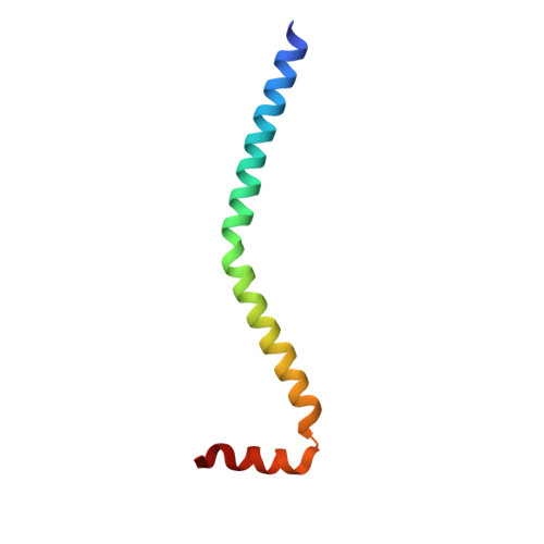
Myosin heavy chain
A, D
69Placopecten magellanicusMutation(s): 0 
UniProt
Find proteins for Q26080 (Placopecten magellanicus)
Explore Q26080 
Go to UniProtKB:  Q26080
Entity Groups  
Sequence Clusters30% Identity50% Identity70% Identity90% Identity95% Identity100% Identity
UniProt GroupQ26080
Sequence Annotations
Expand
  • Reference Sequence
Find similar proteins by:  (by identity cutoff)  |  3D Structure
Entity ID: 2
MoleculeChains Sequence LengthOrganismDetailsImage
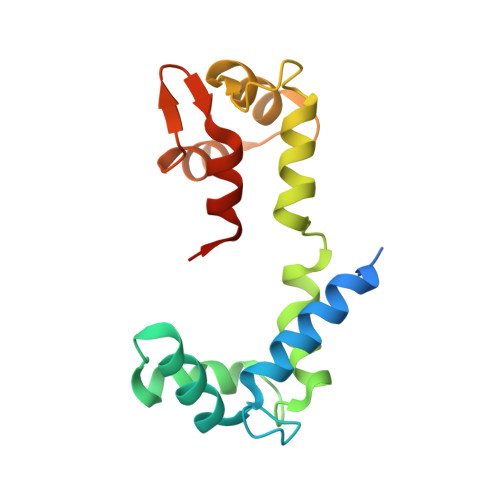
Myosin regulatory light chain
B, E
161Placopecten magellanicusMutation(s): 0 
UniProt
Find proteins for Q26069 (Placopecten magellanicus)
Explore Q26069 
Go to UniProtKB:  Q26069
Entity Groups  
Sequence Clusters30% Identity50% Identity70% Identity90% Identity95% Identity100% Identity
UniProt GroupQ26069
Sequence Annotations
Expand
  • Reference Sequence
Find similar proteins by:  (by identity cutoff)  |  3D Structure
Entity ID: 3
MoleculeChains Sequence LengthOrganismDetailsImage
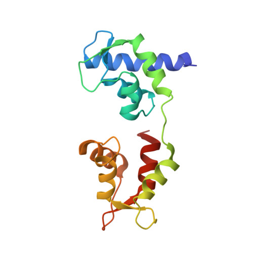
Myosin essential light chain
C, F
156Placopecten magellanicusMutation(s): 0 
UniProt
Find proteins for Q26066 (Placopecten magellanicus)
Explore Q26066 
Go to UniProtKB:  Q26066
Entity Groups  
Sequence Clusters30% Identity50% Identity70% Identity90% Identity95% Identity100% Identity
UniProt GroupQ26066
Sequence Annotations
Expand
  • Reference Sequence
Experimental Data & Validation

Experimental Data

  • Method: X-RAY DIFFRACTION
  • Resolution: 2.39 Å
  • R-Value Free: 0.241 
  • R-Value Work: 0.191 
  • R-Value Observed: 0.195 
  • Space Group: P 1
Unit Cell:
Length ( Å )Angle ( ˚ )
a = 50.811α = 77.33
b = 68.775β = 85.99
c = 79.418γ = 73.65
Software Package:
Software NamePurpose
HKL-2000data collection
PHASERphasing
PHENIXrefinement
HKL-2000data reduction
SCALEPACKdata scaling

Structure Validation

View Full Validation Report


Entry History 

Deposition Data

Revision History  (Full details and data files)

  • Version 1.0: 2011-11-23
    Type: Initial release
  • Version 1.1: 2024-02-28
    Changes: Data collection, Database references, Derived calculations