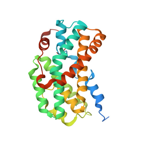
3SF5

Crystal Structure of Helicobacter pylori Urease Accessory Protein UreF/H complex


Experimental Data Snapshot

  • Method: X-RAY DIFFRACTION
  • Resolution: 2.50 Å
  • R-Value Free: 0.217 
  • R-Value Work: 0.173 
  • R-Value Observed: 0.175 

Starting Model: experimental
View more details

wwPDB Validation   3D Report Full Report


This is version 1.2 of the entry. See complete history


Literature

Assembly of preactivation complex for urease maturation in Helicobacter pylori: crystal structure of UreF-UreH protein complex

Fong, Y.H.Wong, H.C.Chuck, C.P.Chen, Y.W.Sun, H.Wong, K.B.

(2011) J Biol Chem 286: 43241-43249

  • DOI: https://doi.org/10.1074/jbc.M111.296830
  • Primary Citation of Related Structures:  
    3O1Q, 3SF5

  • PubMed Abstract: 

    Colonization of Helicobacter pylori in the acidic environment of the human stomach depends on the neutralizing activity of urease. Activation of apo-urease involves carboxylation of lysine 219 and insertion of two nickel ions. In H. pylori, this maturation process involves four urease accessory proteins as follows: UreE, UreF, UreG, and UreH. It is postulated that the apo-urease interacts with UreF, UreG, and UreH to form a pre-activation complex that undergoes GTP-dependent activation of urease. The crystal structure of the UreF-UreH complex reveals conformational changes in two distinct regions of UreF upon complex formation. First, the flexible C-terminal residues of UreF become ordered, forming an extra helix α10 and a loop structure stabilized by hydrogen bonds involving Arg-250. Second, the first turn of helix α2 uncoils to expose a conserved residue, Tyr-48. Substitution of R250A or Y48A in UreF abolishes the formation of the heterotrimeric complex of UreG-UreF-UreH and abolishes urease maturation. Our results suggest that the C-terminal residues and helix α2 of UreF are essential for the recruitment of UreG for the formation of the pre-activation complex. The molecular mass of the UreF-UreH complex determined by static light scattering was 116 ± 2.3 kDa, which is consistent with the quaternary structure of a dimer of heterodimers observed in the crystal structure. Taking advantage of the unique 2-fold symmetry observed in both the crystal structures of H. pylori urease and the UreF-UreH complex, we proposed a topology model of the pre-activation complex for urease maturation.


  • Organizational Affiliation

    Centre for Protein Science and Crystallography, School of Life Sciences, Chinese University of Hong Kong, China, Hong Kong.


Macromolecules
Find similar proteins by:  (by identity cutoff)  |  3D Structure
Entity ID: 1
MoleculeChains Sequence LengthOrganismDetailsImage
Urease accessory protein ureF
A, C
254Helicobacter pyloriMutation(s): 0 
Gene Names: ureF
UniProt
Find proteins for Q09065 (Helicobacter pylori (strain ATCC 700392 / 26695))
Explore Q09065 
Go to UniProtKB:  Q09065
Entity Groups  
Sequence Clusters30% Identity50% Identity70% Identity90% Identity95% Identity100% Identity
UniProt GroupQ09065
Sequence Annotations
Expand
  • Reference Sequence
Find similar proteins by:  (by identity cutoff)  |  3D Structure
Entity ID: 2
MoleculeChains Sequence LengthOrganismDetailsImage
Urease accessory protein ureH
B, D
265Helicobacter pyloriMutation(s): 0 
Gene Names: ureH
UniProt
Find proteins for Q09067 (Helicobacter pylori (strain ATCC 700392 / 26695))
Explore Q09067 
Go to UniProtKB:  Q09067
Entity Groups  
Sequence Clusters30% Identity50% Identity70% Identity90% Identity95% Identity100% Identity
UniProt GroupQ09067
Sequence Annotations
Expand
  • Reference Sequence
Small Molecules
Ligands 3 Unique
IDChains Name / Formula / InChI Key2D Diagram3D Interactions
PEG
Query on PEG

Download Ideal Coordinates CCD File 
E [auth A]
F [auth A]
G [auth A]
H [auth A]
L [auth B]
E [auth A],
F [auth A],
G [auth A],
H [auth A],
L [auth B],
M [auth B],
N [auth C],
O [auth C],
T [auth D]
DI(HYDROXYETHYL)ETHER
C4 H10 O3
MTHSVFCYNBDYFN-UHFFFAOYSA-N
SO4
Query on SO4

Download Ideal Coordinates CCD File 
J [auth B],
K [auth B],
Q [auth C],
R [auth D],
S [auth D]
SULFATE ION
O4 S
QAOWNCQODCNURD-UHFFFAOYSA-L
GOL
Query on GOL

Download Ideal Coordinates CCD File 
I [auth A],
P [auth C]
GLYCEROL
C3 H8 O3
PEDCQBHIVMGVHV-UHFFFAOYSA-N
Experimental Data & Validation

Experimental Data

  • Method: X-RAY DIFFRACTION
  • Resolution: 2.50 Å
  • R-Value Free: 0.217 
  • R-Value Work: 0.173 
  • R-Value Observed: 0.175 
  • Space Group: P 21 21 21
Unit Cell:
Length ( Å )Angle ( ˚ )
a = 70.653α = 90
b = 70.732β = 90
c = 205.499γ = 90
Software Package:
Software NamePurpose
CrystalCleardata collection
PHASERphasing
PHENIXrefinement
XDSdata reduction
SCALAdata scaling

Structure Validation

View Full Validation Report


Entry History 

Deposition Data

Revision History  (Full details and data files)

  • Version 1.0: 2011-11-02
    Type: Initial release
  • Version 1.1: 2012-02-01
    Changes: Database references
  • Version 1.2: 2023-11-01
    Changes: Data collection, Database references, Derived calculations, Refinement description