3SDP

THE 2.1 ANGSTROMS RESOLUTION STRUCTURE OF IRON SUPEROXIDE DISMUTASE FROM PSEUDOMONAS OVALIS


Experimental Data Snapshot

  • Method: X-RAY DIFFRACTION
  • Resolution: 2.10 Å
  • R-Value Observed: 0.232 

wwPDB Validation   3D Report Full Report


This is version 1.3 of the entry. See complete history


Literature

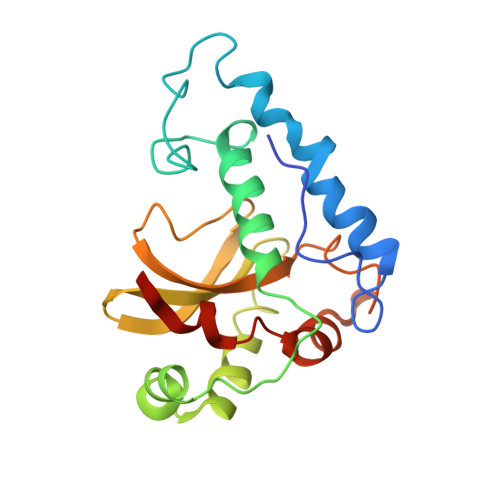
The 2.1-A resolution structure of iron superoxide dismutase from Pseudomonas ovalis.

Stoddard, B.L.Howell, P.L.Ringe, D.Petsko, G.A.

(1990) Biochemistry 29: 8885-8893

  • DOI: https://doi.org/10.1021/bi00490a002
  • Primary Citation of Related Structures:  
    3SDP

  • PubMed Abstract: 

    The 2.1-A resolution crystal structure of native uncomplexed iron superoxide dismutase (EC 1.15.1.1) from Pseudomonas ovalis was solved and refined to a final R factor of 24%. The dimeric structure contains one catalytic iron center per monomer with an asymmetric trigonal-bipyramidal coordination of protein ligands to the metal. Each monomer contains two domains, with the trigonal ligands (histidines 74 and 160; aspartate 156) contributed by the large domain and stabilized by an extended hydrogen-bonded network, including residues from opposing monomers. The axial ligand (histidine 26) is found on the small domain and does not participate extensively in the stabilizing H-bond network. The open axial coordination position of the iron is devoid of bound water molecules or anions. The metal is located 0.5 A out of the plane of the trigonal ligands toward histidine 26, providing a slightly skewed coordination away from the iron binding site. The molecule contains a glutamine residue in the active site which is conserved between all iron enzymes sequenced to data but which is conserved among all manganese SODs at a separate position in the sequence. This residue shows the same structural interactions in both cases, implying that iron and manganese SODs are second-site revertants of one another.


  • Organizational Affiliation

    Department of Chemistry, Massachusetts Institute of Technology, Cambridge 02139.


Macromolecules
Find similar proteins by:  (by identity cutoff)  |  3D Structure
Entity ID: 1
MoleculeChains Sequence LengthOrganismDetailsImage
IRON SUPEROXIDE DISMUTASE
A, B
195Pseudomonas putidaMutation(s): 0 
EC: 1.15.1.1
UniProt
Find proteins for P09223 (Pseudomonas putida)
Explore P09223 
Go to UniProtKB:  P09223
Entity Groups  
Sequence Clusters30% Identity50% Identity70% Identity90% Identity95% Identity100% Identity
UniProt GroupP09223
Sequence Annotations
Expand
  • Reference Sequence
Small Molecules
Ligands 1 Unique
IDChains Name / Formula / InChI Key2D Diagram3D Interactions
FE
Query on FE

Download Ideal Coordinates CCD File 
C [auth A],
D [auth B]
FE (III) ION
Fe
VTLYFUHAOXGGBS-UHFFFAOYSA-N
Experimental Data & Validation

Experimental Data

  • Method: X-RAY DIFFRACTION
  • Resolution: 2.10 Å
  • R-Value Observed: 0.232 
  • Space Group: P 1 21 1
Unit Cell:
Length ( Å )Angle ( ˚ )
a = 81.9α = 90
b = 49.1β = 106.8
c = 61.7γ = 90
Software Package:
Software NamePurpose
X-PLORmodel building
PROLSQrefinement
X-PLORrefinement
X-PLORphasing

Structure Validation

View Full Validation Report


Entry History 

Deposition Data

Revision History  (Full details and data files)

  • Version 1.0: 1993-04-15
    Type: Initial release
  • Version 1.1: 2008-03-03
    Changes: Version format compliance
  • Version 1.2: 2011-07-13
    Changes: Version format compliance
  • Version 1.3: 2024-02-28
    Changes: Data collection, Database references, Derived calculations, Other