3RO1

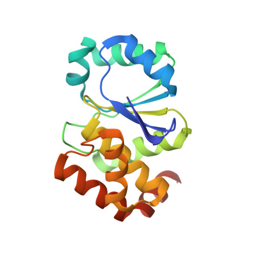
Crystal structure of the complex of the archaeal sulfolobus PTP-fold phosphatase with terpyridine platinum(II)


Experimental Data Snapshot

  • Method: X-RAY DIFFRACTION
  • Resolution: 1.90 Å
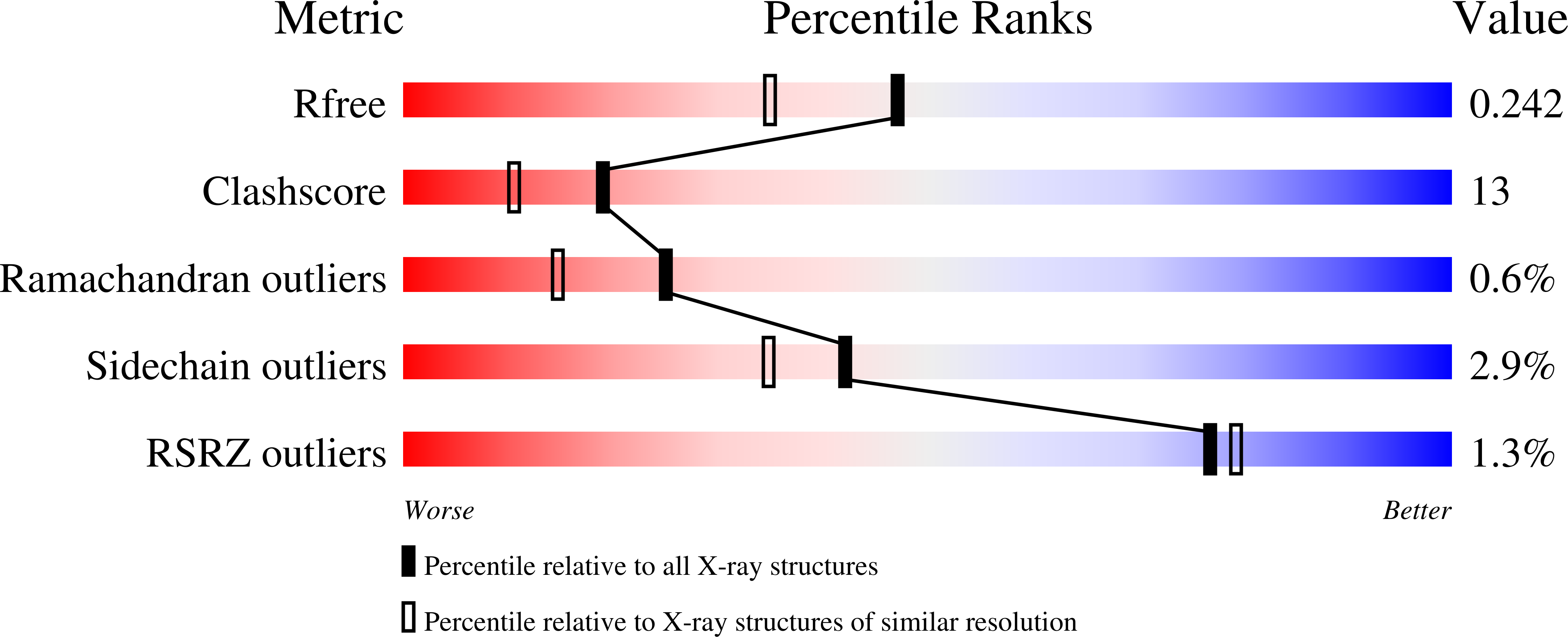
  • R-Value Free: 0.242 
  • R-Value Work: 0.183 
  • R-Value Observed: 0.186 

Starting Model: experimental
View more details

wwPDB Validation   3D Report Full Report


Ligand Structure Quality Assessment 


This is version 1.2 of the entry. See complete history


Literature

Terpyridine platinum(II) complexes inhibit cysteine proteases by binding to active-site cysteine.

Lo, Y.-C.Su, W.-C.Ko, T.-P.Wang, N.-C.Wang, A.H.-J.

(2011) J Biomol Struct Dyn 29: 267-282

  • DOI: https://doi.org/10.1080/073911011010524993
  • Primary Citation of Related Structures:  
    3RNZ, 3RO0, 3RO1

  • PubMed Abstract: 

    Platinum(II) complexes have been demonstrated to form covalent bonds with sulfur-donating ligands (in glutathione, metallothionein and other sulfur-containing biomolecules) or coordination bonds with nitrogen-donating ligands (such as histidine and guanine). To investigate how these compounds interact with cysteine proteases, we chose terpyridine platinum(II) (TP-Pt(II)) complexes as a model system. By using X-ray crystallography, we demonstrated that TP-Pt(II) formed a covalent bond with the catalytic cysteine residue in pyroglutamyl peptidase I. Moreover, by using MALDI (matrix-assisted laser desorption/ionization) and TOF-TOF (time of flight) mass spectrometry, we elucidated that the TP-Pt(II) complex formed a covalent bond with the active-site cysteine residue in two other types of cysteine protease. Taken together, the results unequivocally showed that TP-Pt(II) complexes can selectively bind to the active site of most cysteine proteases. Our findings here can be useful in the design of new anti-cancer, anti-parasite or anti-virus platinum(II) compounds.


  • Organizational Affiliation

    Department and Institute of Pharmacology, National Yang-Ming University, Taipei 112, Taiwan.


Macromolecules
Find similar proteins by:  (by identity cutoff)  |  3D Structure
Entity ID: 1
MoleculeChains Sequence LengthOrganismDetailsImage
protein tyrosine phosphatase161Saccharolobus solfataricusMutation(s): 0 
EC: 3.1.3.48
UniProt
Find proteins for Q97VZ7 (Saccharolobus solfataricus (strain ATCC 35092 / DSM 1617 / JCM 11322 / P2))
Explore Q97VZ7 
Go to UniProtKB:  Q97VZ7
Entity Groups  
Sequence Clusters30% Identity50% Identity70% Identity90% Identity95% Identity100% Identity
UniProt GroupQ97VZ7
Sequence Annotations
Expand
  • Reference Sequence
Small Molecules
Ligands 1 Unique
IDChains Name / Formula / InChI Key2D Diagram3D Interactions
TPT
Query on TPT

Download Ideal Coordinates CCD File 
B [auth A]2,2':6',2''-TERPYRIDINE PLATINUM(II) Chloride
C15 H11 Cl N3 Pt
XTQMQLRZXBEQCS-UHFFFAOYSA-M
Experimental Data & Validation

Experimental Data

  • Method: X-RAY DIFFRACTION
  • Resolution: 1.90 Å
  • R-Value Free: 0.242 
  • R-Value Work: 0.183 
  • R-Value Observed: 0.186 
  • Space Group: P 41
Unit Cell:
Length ( Å )Angle ( ˚ )
a = 72.756α = 90
b = 72.756β = 90
c = 32.302γ = 90
Software Package:
Software NamePurpose
HKL-2000data collection
CNSrefinement
HKL-2000data reduction
HKL-2000data scaling
CNSphasing

Structure Validation

View Full Validation Report


Ligand Structure Quality Assessment 


Entry History 

Deposition Data

Revision History  (Full details and data files)

  • Version 1.0: 2011-10-05
    Type: Initial release
  • Version 1.1: 2019-10-16
    Changes: Data collection, Database references
  • Version 1.2: 2023-11-01
    Changes: Data collection, Database references, Derived calculations, Refinement description