3RNY

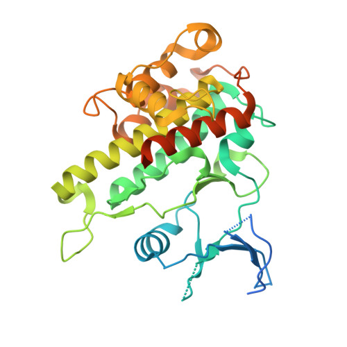
Crystal structure of human RSK1 C-terminal kinase domain


Experimental Data Snapshot

  • Method: X-RAY DIFFRACTION
  • Resolution: 2.70 Å
  • R-Value Free: 0.238 
  • R-Value Work: 0.188 
  • R-Value Observed: 0.191 

Starting Model: experimental
View more details

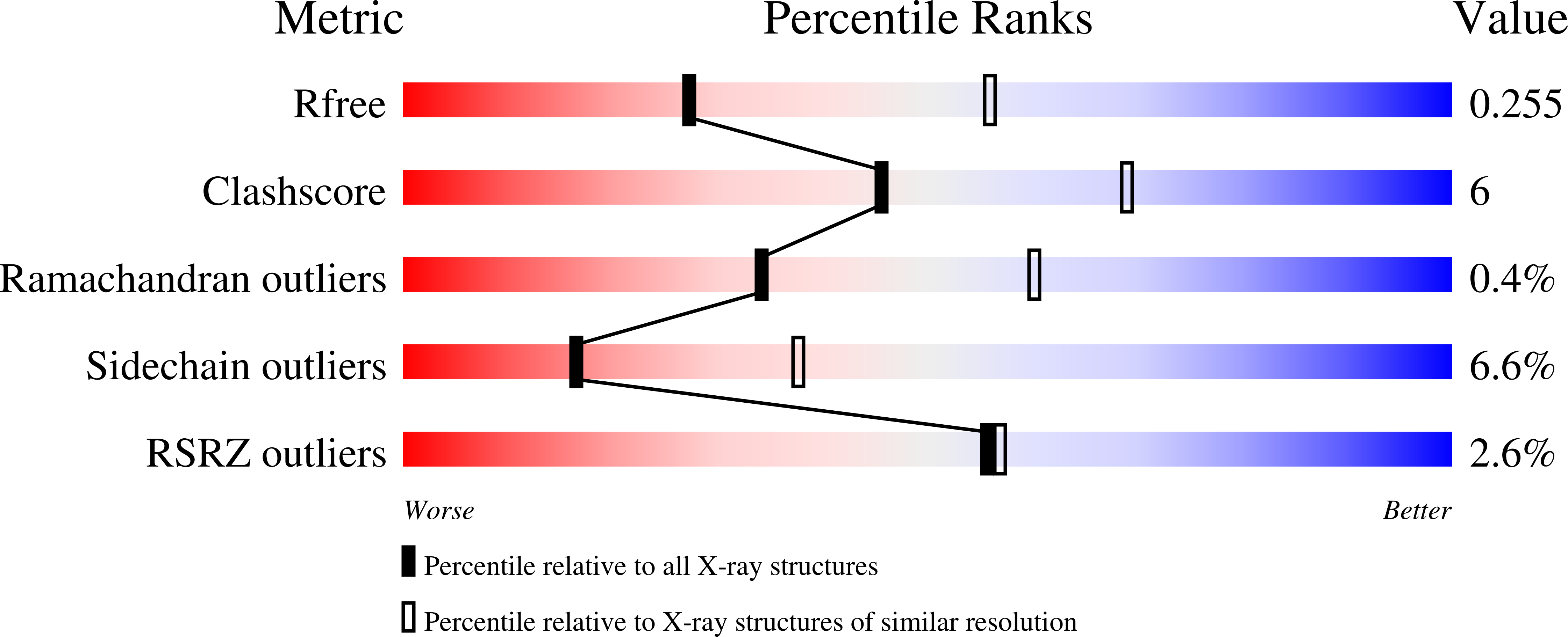
wwPDB Validation   3D Report Full Report


This is version 1.2 of the entry. See complete history


Literature

Structural basis for the autoinhibition of the C-terminal kinase domain of human RSK1.

Li, D.Fu, T.M.Nan, J.Liu, C.Li, L.F.Su, X.D.

(2012) Acta Crystallogr D Biol Crystallogr 68: 680-685

  • DOI: https://doi.org/10.1107/S0907444912007457
  • Primary Citation of Related Structures:  
    3RNY

  • PubMed Abstract: 

    p90 ribosomal S6 kinases (RSKs) respond to various mitogen stimuli and comprise two distinct protein kinase domains. The C-terminal kinase domain (CTKD) receives signal from ERK1/2 and adopts an autoinhibitory mechanism. Here, the crystal structure of human RSK1 CTKD is reported at 2.7 Å resolution. The structure shows a standard kinase fold, with the catalytic residues in the ATP-binding cleft orientated in optimal conformations for phosphotransfer. The inactivation of the CTKD is conferred by an extra α-helix (αL), which occupies the substrate-binding groove. In combination with previous knowledge, this structure indicates that activation of RSK1 involves the removal of αL from the substrate-binding groove induced by ERK1/2 phosphorylation.


  • Organizational Affiliation

    State Key Laboratory of Protein and Plant Gene Research, School of Life Sciences, Peking University, Beijing 100871, People's Republic of China.


Macromolecules
Find similar proteins by:  (by identity cutoff)  |  3D Structure
Entity ID: 1
MoleculeChains Sequence LengthOrganismDetailsImage
Ribosomal protein S6 kinase alpha-1
A, B
346Homo sapiensMutation(s): 0 
Gene Names: RPS6KA1MAPKAPK1ARSK1
EC: 2.7.11.1
UniProt & NIH Common Fund Data Resources
Find proteins for Q15418 (Homo sapiens)
Explore Q15418 
Go to UniProtKB:  Q15418
PHAROS:  Q15418
GTEx:  ENSG00000117676 
Entity Groups  
Sequence Clusters30% Identity50% Identity70% Identity90% Identity95% Identity100% Identity
UniProt GroupQ15418
Sequence Annotations
Expand
  • Reference Sequence
Experimental Data & Validation

Experimental Data

  • Method: X-RAY DIFFRACTION
  • Resolution: 2.70 Å
  • R-Value Free: 0.238 
  • R-Value Work: 0.188 
  • R-Value Observed: 0.191 
  • Space Group: P 1 21 1
Unit Cell:
Length ( Å )Angle ( ˚ )
a = 39.86α = 90
b = 143.47β = 95.77
c = 59.92γ = 90
Software Package:
Software NamePurpose
BUSTER-TNTrefinement
PDB_EXTRACTdata extraction
HKL-2000data collection
HKL-2000data reduction
HKL-2000data scaling
MOLREPphasing
BUSTERrefinement

Structure Validation

View Full Validation Report


Entry History 

Deposition Data

Revision History  (Full details and data files)

  • Version 1.0: 2012-04-25
    Type: Initial release
  • Version 1.1: 2012-08-29
    Changes: Database references
  • Version 1.2: 2023-09-13
    Changes: Data collection, Database references, Derived calculations, Refinement description