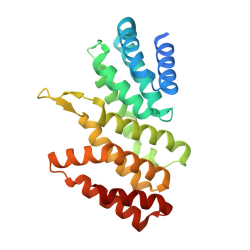
3RK6

Crystal structure of the middle domain of human Paip1


Experimental Data Snapshot

  • Method: X-RAY DIFFRACTION
  • Resolution: 2.00 Å
  • R-Value Free: 0.244 
  • R-Value Work: 0.190 
  • R-Value Observed: 0.192 

wwPDB Validation   3D Report Full Report


This is version 1.3 of the entry. See complete history


Literature

Crystal structure of the middle domain of human poly(A)-binding protein-interacting protein 1.

Lei, J.Mesters, J.R.Brunn, A.Hilgenfeld, R.

(2011) Biochem Biophys Res Commun 408: 680-685

  • DOI: https://doi.org/10.1016/j.bbrc.2011.04.088
  • Primary Citation of Related Structures:  
    3RK6

  • PubMed Abstract: 

    In eukaryotes, the poly(A)-binding protein (PABP) is one of the important factors for initiation of messenger RNA translation. PABP activity is regulated by the PABP-interacting proteins (Paips), which include Paip1, Paip2A, and Paip2B. Human Paip1 has three different isoforms. Here, we report the crystal structure of the middle domain of Paip1 isoform 2 (Paip1M) as determined by single-wavelength anomalous dispersion phasing. The structure reveals a crescent-shaped domain consisting of 10 α-helices and two antiparallel β-strands forming a β-hairpin. The 10 α-helices are arranged as five HEAT repeats which form a double layer of α helices with a convex and a concave surface. Despite low sequence identity, the overall fold of Paip1M is similar to the middle domain of human eIF4GII and yeast eIF4GI. Moreover, the amino-acid sequence motif and the local structure of eIF4G involved in binding of eIF4A, are conserved in Paip1. The structure reported here is the first of a member of the Paip family, thereby filling a gap in our understanding of initiation of eukaryotic mRNA translation in three dimensions.


  • Organizational Affiliation

    Institute of Biochemistry, Center for Structural and Cell Biology in Medicine, University of Lübeck, Ratzeburger Allee 160, Lübeck, Germany.


Macromolecules
Find similar proteins by:  (by identity cutoff)  |  3D Structure
Entity ID: 1
MoleculeChains Sequence LengthOrganismDetailsImage
Polyadenylate-binding protein-interacting protein 1
A, B
234Homo sapiensMutation(s): 0 
Gene Names: PAIP1
UniProt & NIH Common Fund Data Resources
Find proteins for Q9H074 (Homo sapiens)
Explore Q9H074 
Go to UniProtKB:  Q9H074
PHAROS:  Q9H074
GTEx:  ENSG00000172239 
Entity Groups  
Sequence Clusters30% Identity50% Identity70% Identity90% Identity95% Identity100% Identity
UniProt GroupQ9H074
Sequence Annotations
Expand
  • Reference Sequence
Experimental Data & Validation

Experimental Data

  • Method: X-RAY DIFFRACTION
  • Resolution: 2.00 Å
  • R-Value Free: 0.244 
  • R-Value Work: 0.190 
  • R-Value Observed: 0.192 
  • Space Group: P 1 21 1
Unit Cell:
Length ( Å )Angle ( ˚ )
a = 58.61α = 90
b = 76.05β = 96.47
c = 62.42γ = 90
Software Package:
Software NamePurpose
MOSFLMdata reduction
SCALAdata scaling
SHELXphasing
REFMACrefinement
PDB_EXTRACTdata extraction
MAR345dtbdata collection
SHELXCDphasing
SHELXEmodel building

Structure Validation

View Full Validation Report


Entry History 

Deposition Data

Revision History  (Full details and data files)

  • Version 1.0: 2011-05-18
    Type: Initial release
  • Version 1.1: 2011-07-13
    Changes: Version format compliance
  • Version 1.2: 2017-11-08
    Changes: Refinement description
  • Version 1.3: 2024-02-28
    Changes: Data collection, Database references