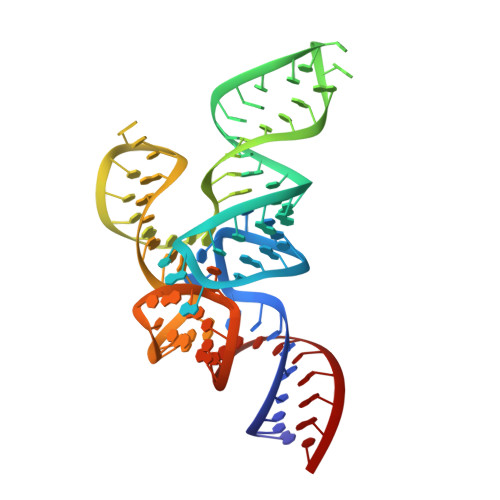
Crystal structure analysis reveals functional flexibility in the selenocysteine-specific tRNA from mouse.
Ganichkin, O.M., Anedchenko, E.A., Wahl, M.C.(2011) PLoS One 6: e20032-e20032
- PubMed: 21629646
- DOI: https://doi.org/10.1371/journal.pone.0020032
- Primary Citation of Related Structures:
3RG5 - PubMed Abstract:
Selenocysteine tRNAs (tRNA(Sec)) exhibit a number of unique identity elements that are recognized specifically by proteins of the selenocysteine biosynthetic pathways and decoding machineries. Presently, these identity elements and the mechanisms by which they are interpreted by tRNA(Sec)-interacting factors are incompletely understood. We applied rational mutagenesis to obtain well diffracting crystals of murine tRNA(Sec). tRNA(Sec) lacking the single-stranded 3'-acceptor end ((ΔGCCA)RNA(Sec)) yielded a crystal structure at 2.0 Å resolution. The global structure of (ΔGCCA)RNA(Sec) resembles the structure of human tRNA(Sec) determined at 3.1 Å resolution. Structural comparisons revealed flexible regions in tRNA(Sec) used for induced fit binding to selenophosphate synthetase. Water molecules located in the present structure were involved in the stabilization of two alternative conformations of the anticodon stem-loop. Modeling of a 2'-O-methylated ribose at position U34 of the anticodon loop as found in a sub-population of tRNA(Sec)in vivo showed how this modification favors an anticodon loop conformation that is functional during decoding on the ribosome. Soaking of crystals in Mn(2+)-containing buffer revealed eight potential divalent metal ion binding sites but the located metal ions did not significantly stabilize specific structural features of tRNA(Sec). We provide the most highly resolved structure of a tRNA(Sec) molecule to date and assessed the influence of water molecules and metal ions on the molecule's conformation and dynamics. Our results suggest how conformational changes of tRNA(Sec) support its interaction with proteins.
Organizational Affiliation:
Abteilung Strukturbiochemie, Freie Universität Berlin, Berlin, Germany.


