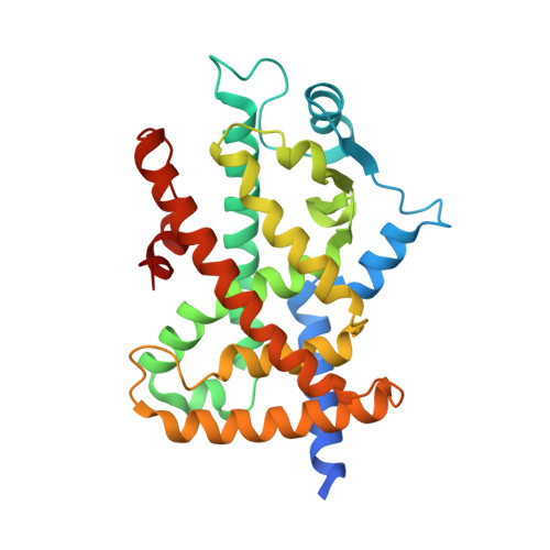
3R8I

Crystal Structure of PPARgamma with an achiral ureidofibrate derivative (RT86)


Experimental Data Snapshot

  • Method: X-RAY DIFFRACTION
  • Resolution: 2.30 Å
  • R-Value Free: 0.285 
  • R-Value Work: 0.252 
  • R-Value Observed: 0.252 

Starting Model: experimental
View more details

wwPDB Validation   3D Report Full Report


Ligand Structure Quality Assessment 


This is version 1.2 of the entry. See complete history


Literature

Synthesis, characterization and biological evaluation of ureidofibrate-like derivatives endowed with peroxisome proliferator-activated receptor activity.

Porcelli, L.Gilardi, F.Laghezza, A.Piemontese, L.Mitro, N.Azzariti, A.Altieri, F.Cervoni, L.Fracchiolla, G.Giudici, M.Guerrini, U.Lavecchia, A.Montanari, R.Di Giovanni, C.Paradiso, A.Pochetti, G.Simone, G.M.Tortorella, P.Crestani, M.Loiodice, F.

(2012) J Med Chem 55: 37-54

  • DOI: https://doi.org/10.1021/jm201306q
  • Primary Citation of Related Structures:  
    3R8I

  • PubMed Abstract: 

    A series of ureidofibrate-like derivatives was prepared and assayed for their PPAR functional activity. A calorimetric approach was used to characterize PPARγ-ligand interactions, and docking experiments and X-ray studies were performed to explain the observed potency and efficacy. R-1 and S-1 were selected to evaluate several aspects of their biological activity. In an adipogenic assay, both enantiomers increased the expression of PPARγ target genes and promoted the differentiation of 3T3-L1 fibroblasts to adipocytes. In vivo administration of these compounds to insulin resistant C57Bl/6J mice fed a high fat diet reduced visceral fat content and body weight. Examination of different metabolic parameters showed that R-1 and S-1 are insulin sensitizers. Notably, they also enhanced the expression of hepatic PPARα target genes indicating that their in vivo effects stemmed from an activation of both PPARα and γ. Finally, the capability of R-1 and S-1 to inhibit cellular proliferation in colon cancer cell lines was also evaluated.


  • Organizational Affiliation

    Laboratorio di Oncologia Sperimentale Clinica, Istituto Tumori "Giovanni Paolo II", 70124 Bari, Italy.


Macromolecules
Find similar proteins by:  (by identity cutoff)  |  3D Structure
Entity ID: 1
MoleculeChains Sequence LengthOrganismDetailsImage
Peroxisome proliferator-activated receptor gamma
A, B
287Homo sapiensMutation(s): 0 
Gene Names: PPARGNR1C3
UniProt & NIH Common Fund Data Resources
Find proteins for P37231 (Homo sapiens)
Explore P37231 
Go to UniProtKB:  P37231
PHAROS:  P37231
GTEx:  ENSG00000132170 
Entity Groups  
Sequence Clusters30% Identity50% Identity70% Identity90% Identity95% Identity100% Identity
UniProt GroupP37231
Sequence Annotations
Expand
  • Reference Sequence
Small Molecules
Ligands 1 Unique
IDChains Name / Formula / InChI Key2D Diagram3D Interactions
XCX
Query on XCX

Download Ideal Coordinates CCD File 
C [auth A]2-(4-{2-[1,3-benzoxazol-2-yl(heptyl)amino]ethyl}phenoxy)-2-methylpropanoic acid
C26 H34 N2 O4
MMVUCPZZXMPIPE-UHFFFAOYSA-N
Binding Affinity Annotations 
IDSourceBinding Affinity
XCX BindingDB:  3R8I Kd: 4500 (nM) from 1 assay(s)
EC50: 110 (nM) from 1 assay(s)
PDBBind:  3R8I Kd: 4500 (nM) from 1 assay(s)
Experimental Data & Validation

Experimental Data

  • Method: X-RAY DIFFRACTION
  • Resolution: 2.30 Å
  • R-Value Free: 0.285 
  • R-Value Work: 0.252 
  • R-Value Observed: 0.252 
  • Space Group: C 1 2 1
Unit Cell:
Length ( Å )Angle ( ˚ )
a = 93.589α = 90
b = 62.398β = 102.11
c = 118.392γ = 90
Software Package:
Software NamePurpose
ADSCdata collection
AMoREphasing
CNSrefinement
MOSFLMdata reduction
SCALAdata scaling

Structure Validation

View Full Validation Report


Ligand Structure Quality Assessment 


Entry History 

Deposition Data

Revision History  (Full details and data files)

  • Version 1.0: 2012-01-11
    Type: Initial release
  • Version 1.1: 2012-04-25
    Changes: Database references
  • Version 1.2: 2023-09-13
    Changes: Data collection, Database references, Derived calculations, Refinement description