Identification of the PDZ3 Domain of the Adaptor Protein PDZK1 as a Second, Physiologically Functional Binding Site for the C Terminus of the High Density Lipoprotein Receptor Scavenger Receptor Class B Type I.
Kocher, O., Birrane, G., Yesilaltay, A., Shechter, S., Pal, R., Daniels, K., Krieger, M.(2011) J Biol Chem 286: 25171-25186
- PubMed: 21602281
- DOI: https://doi.org/10.1074/jbc.M111.242362
- Primary Citation of Related Structures:
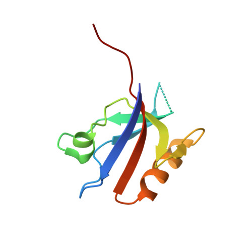
3R68, 3R69 - PubMed Abstract:
The normal expression, cell surface localization, and function of the murine high density lipoprotein receptor scavenger receptor class B type I (SR-BI) in hepatocytes in vivo, and thus normal lipoprotein metabolism, depend on its four PDZ domain (PDZ1-PDZ4) containing cytoplasmic adaptor protein PDZK1. Previous studies showed that the C terminus of SR-BI ("target peptide") binds directly to PDZ1 and influences hepatic SR-BI protein expression. Unexpectedly an inactivating mutation in PDZ1 (Tyr(20) → Ala) only partially, rather than completely, suppresses the ability of PDZK1 to control hepatic SR-BI. We used isothermal titration calorimetry to show that PDZ3, but not PDZ2 or PDZ4, can also bind the target peptide (K(d) = 37.0 μm), albeit with ∼10-fold lower affinity than PDZ1. This binding is abrogated by a Tyr(253) → Ala substitution. Comparison of the 1.5-Å resolution crystal structure of PDZ3 with its bound target peptide ((505)QEAKL(509)) to that of peptide-bound PDZ1 indicated fewer target peptide stabilizing atomic interactions (hydrogen bonds and hydrophobic interactions) in PDZ3. A double (Tyr(20) → Ala (PDZ1) + Tyr(253) → Ala (PDZ3)) substitution abrogated all target peptide binding to PDZK1. In vivo hepatic expression of a singly substituted (Tyr(253) → Ala (PDZ3)) PDZK1 transgene (Tg) was able to correct all of the SR-BI-related defects in PDZK1 knock-out mice, whereas the doubly substituted [Tyr(20) → Ala (PDZ1) + Tyr(253) → Ala (PDZ3)]Tg was unable to correct these defects. Thus, we conclude that PDZK1-mediated control of hepatic SR-BI requires direct binding of the SR-BI C terminus to either the PDZ1 or PDZ3 domains, and that binding to both domains simultaneously is not required for PDZK1 control of hepatic SR-BI.
Organizational Affiliation:
Department of Pathology, Harvard Medical School, Boston, Massachusetts 02215, USA. okocher@bidmc.harvard.edu