Tetrahydrodipicolinate N-succinyltransferase and dihydrodipicolinate synthase from Pseudomonas aeruginosa: structure analysis and gene deletion.
Schnell, R., Oehlmann, W., Sandalova, T., Braun, Y., Huck, C., Maringer, M., Singh, M., Schneider, G.(2012) PLoS One 7: e31133-e31133
- PubMed: 22359568
- DOI: https://doi.org/10.1371/journal.pone.0031133
- Primary Citation of Related Structures:
3QZE, 3R5A, 3R5B, 3R5C, 3R5D - PubMed Abstract:
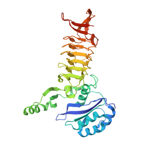
The diaminopimelic acid pathway of lysine biosynthesis has been suggested to provide attractive targets for the development of novel antibacterial drugs. Here we report the characterization of two enzymes from this pathway in the human pathogen Pseudomonas aeruginosa, utilizing structural biology, biochemistry and genetics. We show that tetrahydrodipicolinate N-succinyltransferase (DapD) from P. aeruginosa is specific for the L-stereoisomer of the amino substrate L-2-aminopimelate, and its D-enantiomer acts as a weak inhibitor. The crystal structures of this enzyme with L-2-aminopimelate and D-2-aminopimelate, respectively, reveal that both compounds bind at the same site of the enzyme. Comparison of the binding interactions of these ligands in the enzyme active site suggests misalignment of the amino group of D-2-aminopimelate for nucleophilic attack on the succinate moiety of the co-substrate succinyl-CoA as the structural basis of specificity and inhibition. P. aeruginosa mutants where the dapA gene had been deleted were viable and able to grow in a mouse lung infection model, suggesting that DapA is not an optimal target for drug development against this organism. Structure-based sequence alignments, based on the DapA crystal structure determined to 1.6 Å resolution revealed the presence of two homologues, PA0223 and PA4188, in P. aeruginosa that could substitute for DapA in the P. aeruginosa PAO1ΔdapA mutant. In vitro experiments using recombinant PA0223 protein could however not detect any DapA activity.
Organizational Affiliation:
Department of Medical Biochemistry and Biophysics, Karolinska Institutet, Stockholm, Sweden.


