3OA0

Crystal structure of the WlbA (WbpB) Dehydrogenase from Thermus thermophilus in complex with NAD and UDP-GlcNAcA


Experimental Data Snapshot

  • Method: X-RAY DIFFRACTION
  • Resolution: 2.00 Å
  • R-Value Free: 0.248 
  • R-Value Work: 0.189 
  • R-Value Observed: 0.192 

Starting Model: experimental
View more details

wwPDB Validation   3D Report Full Report


Ligand Structure Quality Assessment 


This is version 1.2 of the entry. See complete history


Literature

Structural and Functional Studies of WlbA: A Dehydrogenase Involved in the Biosynthesis of 2,3-Diacetamido-2,3-dideoxy-d-mannuronic Acid .

Thoden, J.B.Holden, H.M.

(2010) Biochemistry 49: 7939-7948

  • DOI: https://doi.org/10.1021/bi101103s
  • Primary Citation of Related Structures:  
    3O9Z, 3OA0, 3OA2

  • PubMed Abstract: 

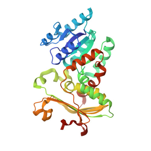
    2,3-Diacetamido-2,3-dideoxy-d-mannuronic acid (ManNAc3NAcA) is an unusual dideoxy sugar first identified nearly 30 years ago in the lipopolysaccharide of Pseudomonas aeruginosa O:3a,d. It has since been observed in other organisms, including Bordetella pertussis, the causative agent of whooping cough. Five enzymes are required for the biosynthesis of UDP-ManNAc3NAcA starting from UDP-N-acetyl-d-glucosamine. Here we describe a structural study of WlbA, the NAD-dependent dehydrogenase that catalyzes the second step in the pathway, namely, the oxidation of the C-3' hydroxyl group on the UDP-linked sugar to a keto moiety and the reduction of NAD(+) to NADH. This enzyme has been shown to use alpha-ketoglutarate as an oxidant to regenerate the oxidized dinucleotide. For this investigation, three different crystal structures were determined: the enzyme with bound NAD(H), the enzyme in a complex with NAD(H) and alpha-ketoglutarate, and the enzyme in a complex with NAD(H) and its substrate (UDP-N-acetyl-d-glucosaminuronic acid). The tetrameric enzyme assumes an unusual quaternary structure with the dinucleotides positioned quite closely to one another. Both alpha-ketoglutarate and the UDP-linked sugar bind in the WlbA active site with their carbon atoms (C-2 and C-3', respectively) abutting the re face of the cofactor. They are positioned approximately 3 A from the nicotinamide C-4. The UDP-linked sugar substrate adopts a highly unusual curved conformation when bound in the WlbA active site cleft. Lys 101 and His 185 most likely play key roles in catalysis.


  • Organizational Affiliation

    Department of Biochemistry, University of Wisconsin, Madison, Wisconsin 53706, USA.


Macromolecules
Find similar proteins by:  (by identity cutoff)  |  3D Structure
Entity ID: 1
MoleculeChains Sequence LengthOrganismDetailsImage
Lipopolysaccharide biosynthesis protein wbpB
A, B, C, D
312Thermus thermophilus HB27Mutation(s): 0 
Gene Names: TT_C0287wbpB
UniProt
Find proteins for Q72KX8 (Thermus thermophilus (strain ATCC BAA-163 / DSM 7039 / HB27))
Explore Q72KX8 
Go to UniProtKB:  Q72KX8
Entity Groups  
Sequence Clusters30% Identity50% Identity70% Identity90% Identity95% Identity100% Identity
UniProt GroupQ72KX8
Sequence Annotations
Expand
  • Reference Sequence
Small Molecules
Ligands 3 Unique
IDChains Name / Formula / InChI Key2D Diagram3D Interactions
NAD
Query on NAD

Download Ideal Coordinates CCD File 
E [auth A],
H [auth B],
K [auth C],
N [auth D]
NICOTINAMIDE-ADENINE-DINUCLEOTIDE
C21 H27 N7 O14 P2
BAWFJGJZGIEFAR-NNYOXOHSSA-N
HP7
Query on HP7

Download Ideal Coordinates CCD File 
F [auth A],
I [auth B],
L [auth C],
O [auth D]
(2S,3S,4R,5R,6R)-5-acetamido-6-[[[(2R,3S,4R,5R)-5-(2,4-dioxopyrimidin-1-yl)-3,4-dihydroxy-oxolan-2-yl]methoxy-hydroxy-phosphoryl]oxy-hydroxy-phosphoryl]oxy-3,4-dihydroxy-oxane-2-carboxylic acid
C17 H25 N3 O18 P2
DZOGQXKQLXAPND-HHKCBAECSA-N
CL
Query on CL

Download Ideal Coordinates CCD File 
G [auth A],
J [auth B],
M [auth C],
P [auth D]
CHLORIDE ION
Cl
VEXZGXHMUGYJMC-UHFFFAOYSA-M
Experimental Data & Validation

Experimental Data

  • Method: X-RAY DIFFRACTION
  • Resolution: 2.00 Å
  • R-Value Free: 0.248 
  • R-Value Work: 0.189 
  • R-Value Observed: 0.192 
  • Space Group: P 1 21 1
Unit Cell:
Length ( Å )Angle ( ˚ )
a = 72.097α = 90
b = 68.32β = 94.51
c = 164.122γ = 90
Software Package:
Software NamePurpose
HKL-3000data collection
PHASERphasing
REFMACrefinement
HKL-3000data reduction
HKL-3000data scaling

Structure Validation

View Full Validation Report


Ligand Structure Quality Assessment 


Entry History 

Deposition Data

Revision History  (Full details and data files)

  • Version 1.0: 2010-08-18
    Type: Initial release
  • Version 1.1: 2011-07-13
    Changes: Version format compliance
  • Version 1.2: 2023-09-06
    Changes: Data collection, Database references, Derived calculations, Refinement description