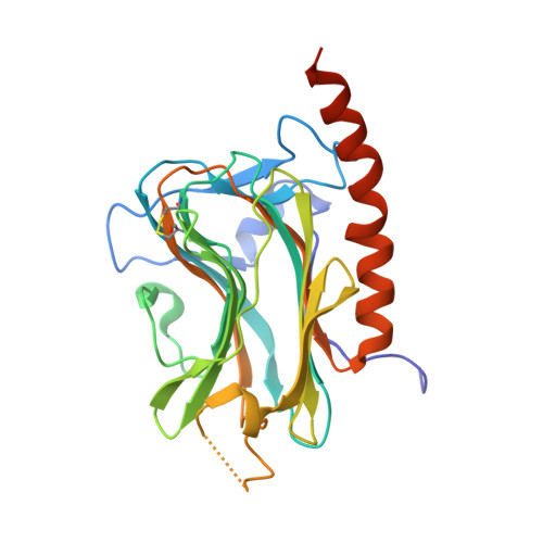
Structural basis of carbohydrate recognition by calreticulin.
Kozlov, G., Pocanschi, C.L., Rosenauer, A., Bastos-Aristizabal, S., Gorelik, A., Williams, D.B., Gehring, K.(2010) J Biol Chem 285: 38612-38620
- PubMed: 20880849
- DOI: https://doi.org/10.1074/jbc.M110.168294
- Primary Citation of Related Structures:
3O0V, 3O0W, 3O0X - PubMed Abstract:
The calnexin cycle is a process by which glycosylated proteins are subjected to folding cycles in the endoplasmic reticulum lumen via binding to the membrane protein calnexin (CNX) or to its soluble homolog calreticulin (CRT). CNX and CRT specifically recognize monoglucosylated Glc(1)Man(9)GlcNAc(2) glycans, but the structural determinants underlying this specificity are unknown. Here, we report a 1.95-Å crystal structure of the CRT lectin domain in complex with the tetrasaccharide α-Glc-(1→3)-α-Man-(1→2)-α-Man-(1→2)-Man. The tetrasaccharide binds to a long channel on CRT formed by a concave β-sheet. All four sugar moieties are engaged in the protein binding via an extensive network of hydrogen bonds and hydrophobic contacts. The structure explains the requirement for glucose at the nonreducing end of the carbohydrate; the oxygen O(2) of glucose perfectly fits to a pocket formed by CRT side chains while forming direct hydrogen bonds with the carbonyl of Gly(124) and the side chain of Lys(111). The structure also explains a requirement for the Cys(105)-Cys(137) disulfide bond in CRT/CNX for efficient carbohydrate binding. The Cys(105)-Cys(137) disulfide bond is involved in intimate contacts with the third and fourth sugar moieties of the Glc(1)Man(3) tetrasaccharide. Finally, the structure rationalizes previous mutagenesis of CRT and lays a structural groundwork for future studies of the role of CNX/CRT in diverse biological pathways.
Organizational Affiliation:
Department of Biochemistry, Groupe de Recherche Axé sur la Structure des Protéines, McGill University, Montréal, Québec H3G 0B1, Canada.


