3NIH

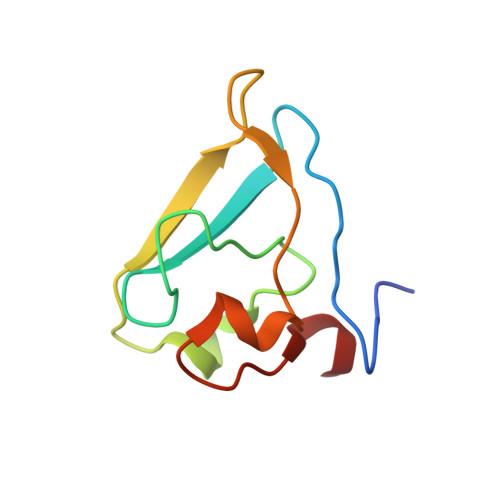
The structure of UBR box (RIAAA)


Experimental Data Snapshot

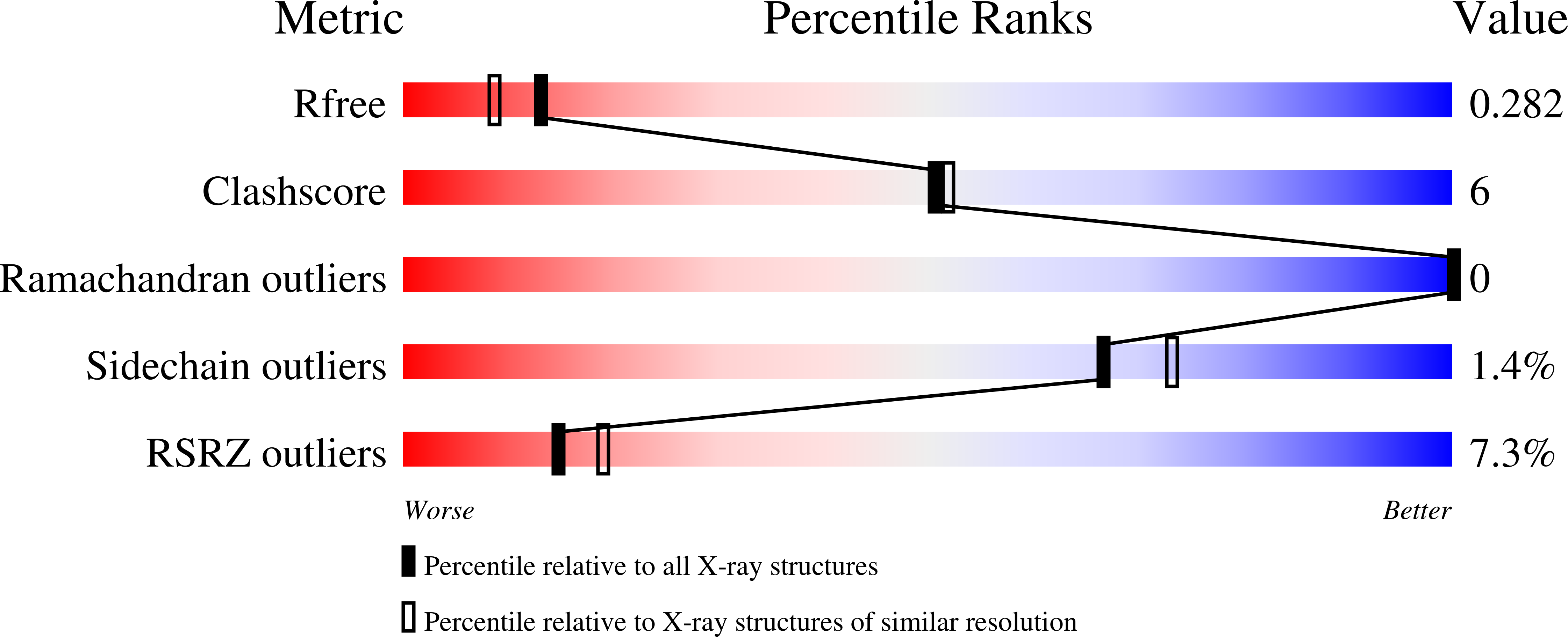
  • Method: X-RAY DIFFRACTION
  • Resolution: 2.10 Å
  • R-Value Free: 0.285 
  • R-Value Work: 0.253 
  • R-Value Observed: 0.255 

Starting Model: experimental
View more details

wwPDB Validation   3D Report Full Report


This is version 1.4 of the entry. See complete history


Literature

Structural basis for the recognition of N-end rule substrates by the UBR box of ubiquitin ligases

Choi, W.S.Jeong, B.-C.Joo, Y.J.Lee, M.-R.Kim, J.Eck, M.J.Song, H.K.

(2010) Nat Struct Mol Biol 17: 1175-1181

  • DOI: https://doi.org/10.1038/nsmb.1907
  • Primary Citation of Related Structures:  
    3NIH, 3NII, 3NIJ, 3NIK, 3NIL, 3NIM, 3NIN, 3NIS, 3NIT

  • PubMed Abstract: 

    The N-end rule pathway is a regulated proteolytic system that targets proteins containing destabilizing N-terminal residues (N-degrons) for ubiquitination and proteasomal degradation in eukaryotes. The N-degrons of type 1 substrates contain an N-terminal basic residue that is recognized by the UBR box domain of the E3 ubiquitin ligase UBR1. We describe structures of the UBR box of Saccharomyces cerevisiae UBR1 alone and in complex with N-degron peptides, including that of the cohesin subunit Scc1, which is cleaved and targeted for degradation at the metaphase-anaphase transition. The structures reveal a previously unknown protein fold that is stabilized by a novel binuclear zinc center. N-terminal arginine, lysine or histidine side chains of the N-degron are coordinated in a multispecific binding pocket. Unexpectedly, the structures together with our in vitro biochemical and in vivo pulse-chase analyses reveal a previously unknown modulation of binding specificity by the residue at position 2 of the N-degron.


  • Organizational Affiliation

    School of Life Sciences and Biotechnology, Korea University, Anam-Dong, Korea.


Macromolecules
Find similar proteins by:  (by identity cutoff)  |  3D Structure
Entity ID: 1
MoleculeChains Sequence LengthOrganismDetailsImage
E3 ubiquitin-protein ligase UBR182Saccharomyces cerevisiaeMutation(s): 0 
EC: 2.3.2.27
UniProt
Find proteins for P19812 (Saccharomyces cerevisiae (strain ATCC 204508 / S288c))
Explore P19812 
Go to UniProtKB:  P19812
Entity Groups  
Sequence Clusters30% Identity50% Identity70% Identity90% Identity95% Identity100% Identity
UniProt GroupP19812
Sequence Annotations
Expand
  • Reference Sequence

Find similar proteins by:  Sequence   |   3D Structure  

Entity ID: 2
MoleculeChains Sequence LengthOrganismDetailsImage
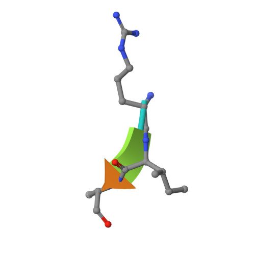
Peptide RIAAA5N/AMutation(s): 0 
Sequence Annotations
Expand
  • Reference Sequence
Experimental Data & Validation

Experimental Data

  • Method: X-RAY DIFFRACTION
  • Resolution: 2.10 Å
  • R-Value Free: 0.285 
  • R-Value Work: 0.253 
  • R-Value Observed: 0.255 
  • Space Group: P 61 2 2
Unit Cell:
Length ( Å )Angle ( ˚ )
a = 58.274α = 90
b = 58.274β = 90
c = 110.999γ = 120
Software Package:
Software NamePurpose
DENZOdata reduction
SCALEPACKdata scaling
REFMACrefinement
PDB_EXTRACTdata extraction
HKL-2000data collection
HKL-2000data reduction
HKL-2000data scaling
MOLREPphasing

Structure Validation

View Full Validation Report


Entry History 

Deposition Data

Revision History  (Full details and data files)

  • Version 1.0: 2010-09-15
    Type: Initial release
  • Version 1.1: 2011-07-13
    Changes: Version format compliance
  • Version 1.2: 2017-11-08
    Changes: Refinement description
  • Version 1.3: 2023-11-01
    Changes: Data collection, Database references, Derived calculations, Refinement description
  • Version 1.4: 2024-10-30
    Changes: Structure summary