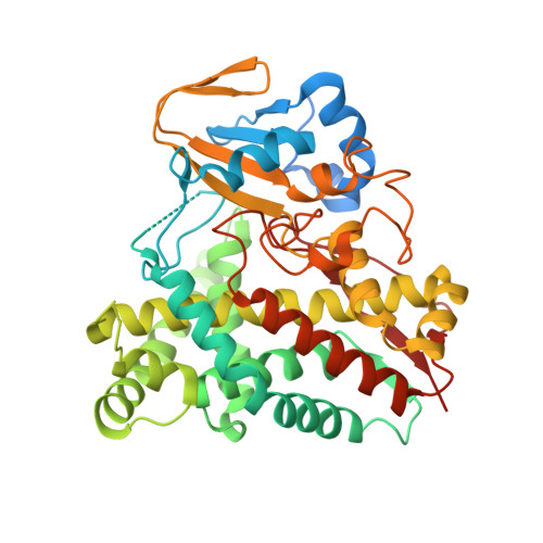
Structural and biochemical characterization of the cytochrome P450 CypX (CYP134A1) from Bacillus subtilis: a cyclo-L-leucyl-L-leucyl dipeptide oxidase.
Cryle, M.J., Bell, S.G., Schlichting, I.(2010) Biochemistry 49: 7282-7296
- PubMed: 20690619
- DOI: https://doi.org/10.1021/bi100910y
- Primary Citation of Related Structures:
3NC3, 3NC5, 3NC6, 3NC7 - PubMed Abstract:
Cytochrome P450 CypX (CYP134A1), isolated from Bacillus subtilis, has previously been implicated in the three-step oxidative transformation of the diketopiperazine cyclo-l-leucyl-l-leucyl into pulcherriminic acid, a precursor of the extracellular iron chelate pulcherrimin. In this study, we present the first experimental data relating to CYP134A1, where we show that CYP134A1 binds cyclo-l-leucyl-l-leucyl with an affinity of 24.5 +/- 0.5 muM. Structurally related diketopiperazines sharing similar alkyl side chains to cyclo-l-leucyl-l-leucyl also bind to CYP134A1 with comparable affinity. CYP134A1 is capable of catalyzing the in vitro oxidation of diketopiperazine substrates when supported with several alternate electron transfer partner systems. Products containing one additional oxygen atom and which are intermediate products of the expected pulcherriminic acid were identified by GCMS. The oxidation of related diketopiperazines reveals that different oxidative pathways exist for CYP134A1-catalyzed diketopiperazine oxidation. The crystal structure of CYP134A1 has been determined to 2.7 A resolution in the absence of substrate and in the presence of bound phenylimidazole ligands to 3.1 and 3.2 A resolution. The active site is dominated by hydrophobic residues and contains an unusual proline residue in place of the normally conserved alcohol residue that typically plays an important role in oxygen activation. The B-B(2) substrate recognition loop, which forms part of the active site, shows considerable flexibility and was found in both open and closed conformations in different structures. These results represent the first insights into the structural and biochemical basis underlying the multistep oxidation catalyzed by CYP134A1.
Organizational Affiliation:
Department of Biomolecular Mechanisms, Max Planck Institute for Medical Research, Jahnstrasse 29, 69120 Heidelberg, Germany. Max.Cryle@mpimf-heidelberg.mpg.de


