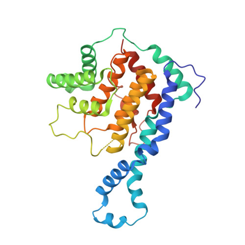
3N3U

Crystal Structure of IbpAFic2


Experimental Data Snapshot

  • Method: X-RAY DIFFRACTION
  • Resolution: 1.85 Å
  • R-Value Free: 0.242 
  • R-Value Work: 0.212 
  • R-Value Observed: 0.213 

wwPDB Validation   3D Report Full Report


This is version 1.4 of the entry. See complete history


Literature

Structural basis of Fic-mediated adenylylation.

Xiao, J.Worby, C.A.Mattoo, S.Sankaran, B.Dixon, J.E.

(2010) Nat Struct Mol Biol 17: 1004-1010

  • DOI: https://doi.org/10.1038/nsmb.1867
  • Primary Citation of Related Structures:  
    3N3U, 4ITR

  • PubMed Abstract: 

    The Fic family of adenylyltransferases, defined by a core HPFx(D/E)GN(G/K)R motif, consists of over 2,700 proteins found in organisms from bacteria to humans. The immunoglobulin-binding protein A (IbpA) from the bacterial pathogen Histophilus somni contains two Fic domains that adenylylate the switch1 tyrosine residue of Rho-family GTPases, allowing the bacteria to subvert host defenses. Here we present the structure of the second Fic domain of IbpA (IbpAFic2) in complex with its substrate, Cdc42. IbpAFic2-bound Cdc42 mimics the GDI-bound state of Rho GTPases, with both its switch1 and switch2 regions gripped by IbpAFic2. Mutations disrupting the IbpAFic2-Cdc42 interface impair adenylylation and cytotoxicity. Notably, the switch1 tyrosine of Cdc42 is adenylylated in the structure, providing the first structural view for this post-translational modification. We also show that the nucleotide-binding mechanism is conserved among Fic proteins and propose a catalytic mechanism for this recently discovered family of enzymes.


  • Organizational Affiliation

    Department of Pharmacology, Howard Hughes Medical Institute, University of California, San Diego, La Jolla, California, USA.


Macromolecules
Find similar proteins by:  (by identity cutoff)  |  3D Structure
Entity ID: 1
MoleculeChains Sequence LengthOrganismDetailsImage
Adenosine monophosphate-protein transferase ibpA299Histophilus somni 2336Mutation(s): 0 
Gene Names: HSM_1489ibpAp76
EC: 2.7.7.1 (PDB Primary Data), 3.4.22 (UniProt), 2.7.7.108 (UniProt)
UniProt
Find proteins for Q06277 (Histophilus somni (strain 2336))
Explore Q06277 
Go to UniProtKB:  Q06277
Entity Groups  
Sequence Clusters30% Identity50% Identity70% Identity90% Identity95% Identity100% Identity
UniProt GroupQ06277
Sequence Annotations
Expand
  • Reference Sequence
Experimental Data & Validation

Experimental Data

  • Method: X-RAY DIFFRACTION
  • Resolution: 1.85 Å
  • R-Value Free: 0.242 
  • R-Value Work: 0.212 
  • R-Value Observed: 0.213 
  • Space Group: P 31 2 1
Unit Cell:
Length ( Å )Angle ( ˚ )
a = 64.38α = 90
b = 64.38β = 90
c = 149.689γ = 120
Software Package:
Software NamePurpose
DENZOdata reduction
SCALEPACKdata scaling
PHENIXrefinement
PDB_EXTRACTdata extraction
AutoSolphasing

Structure Validation

View Full Validation Report


Entry History 

Deposition Data

  • Released Date: 2010-07-14 
  • Deposition Author(s): Xiao, J.

Revision History  (Full details and data files)

  • Version 1.0: 2010-07-14
    Type: Initial release
  • Version 1.1: 2011-07-13
    Changes: Version format compliance
  • Version 1.2: 2011-11-16
    Changes: Atomic model
  • Version 1.3: 2017-11-08
    Changes: Refinement description
  • Version 1.4: 2024-02-21
    Changes: Data collection, Database references, Derived calculations