3MKS

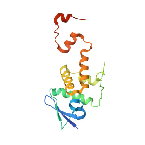
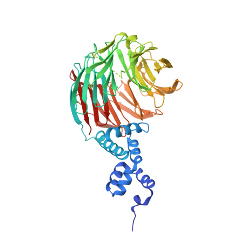
Crystal Structure of yeast Cdc4/Skp1 in complex with an allosteric inhibitor SCF-I2


Experimental Data Snapshot

  • Method: X-RAY DIFFRACTION
  • Resolution: 2.60 Å
  • R-Value Free: 0.266 
  • R-Value Work: 0.211 
  • R-Value Observed: 0.213 

Starting Model: experimental
View more details

wwPDB Validation   3D Report Full Report


Ligand Structure Quality Assessment 


This is version 1.3 of the entry. See complete history


Literature

An allosteric inhibitor of substrate recognition by the SCF(Cdc4) ubiquitin ligase.

Orlicky, S.Tang, X.Neduva, V.Elowe, N.Brown, E.D.Sicheri, F.Tyers, M.

(2010) Nat Biotechnol 28: 733-737

  • DOI: https://doi.org/10.1038/nbt.1646
  • Primary Citation of Related Structures:  
    3MKS

  • PubMed Abstract: 

    The specificity of SCF ubiquitin ligase-mediated protein degradation is determined by F-box proteins. We identified a biplanar dicarboxylic acid compound, called SCF-I2, as an inhibitor of substrate recognition by the yeast F-box protein Cdc4 using a fluorescence polarization screen to monitor the displacement of a fluorescein-labeled phosphodegron peptide. SCF-I2 inhibits the binding and ubiquitination of full-length phosphorylated substrates by SCF(Cdc4). A co-crystal structure reveals that SCF-I2 inserts itself between the beta-strands of blades 5 and 6 of the WD40 propeller domain of Cdc4 at a site that is 25 A away from the substrate binding site. Long-range transmission of SCF-I2 interactions distorts the substrate binding pocket and impedes recognition of key determinants in the Cdc4 phosphodegron. Mutation of the SCF-I2 binding site abrogates its inhibitory effect and explains specificity in the allosteric inhibition mechanism. Mammalian WD40 domain proteins may exhibit similar allosteric responsiveness and hence represent an extensive class of druggable target.


  • Organizational Affiliation

    Center for Systems Biology, Samuel Lunenfeld Research Institute, Mount Sinai Hospital, Toronto, Canada.


Macromolecules
Find similar proteins by:  (by identity cutoff)  |  3D Structure
Entity ID: 1
MoleculeChains Sequence LengthOrganismDetailsImage
Suppressor of kinetochore protein 1
A, C
169Saccharomyces cerevisiaeMutation(s): 0 
Gene Names: SKP1CBF3DYDR328CD9798.14
UniProt
Find proteins for P52286 (Saccharomyces cerevisiae (strain ATCC 204508 / S288c))
Explore P52286 
Go to UniProtKB:  P52286
Entity Groups  
Sequence Clusters30% Identity50% Identity70% Identity90% Identity95% Identity100% Identity
UniProt GroupP52286
Sequence Annotations
Expand
  • Reference Sequence
Find similar proteins by:  (by identity cutoff)  |  3D Structure
Entity ID: 2
MoleculeChains Sequence LengthOrganismDetailsImage
Cell division control protein 4
B, D
464Saccharomyces cerevisiaeMutation(s): 0 
Gene Names: CDC4YFL009W
UniProt
Find proteins for P07834 (Saccharomyces cerevisiae (strain ATCC 204508 / S288c))
Explore P07834 
Go to UniProtKB:  P07834
Entity Groups  
Sequence Clusters30% Identity50% Identity70% Identity90% Identity95% Identity100% Identity
UniProt GroupP07834
Sequence Annotations
Expand
  • Reference Sequence
Small Molecules
Ligands 3 Unique
IDChains Name / Formula / InChI Key2D Diagram3D Interactions
C1C
Query on C1C

Download Ideal Coordinates CCD File 
J [auth D]1,1'-binaphthalene-2,2'-dicarboxylic acid
C22 H14 O4
YDZNRNHKJQTGCG-UHFFFAOYSA-N
SO4
Query on SO4

Download Ideal Coordinates CCD File 
E [auth B]
F [auth B]
G [auth B]
H [auth B]
I [auth B]
E [auth B],
F [auth B],
G [auth B],
H [auth B],
I [auth B],
K [auth D],
L [auth D],
M [auth D],
N [auth D],
O [auth D],
P [auth D]
SULFATE ION
O4 S
QAOWNCQODCNURD-UHFFFAOYSA-L
GOL
Query on GOL

Download Ideal Coordinates CCD File 
Q [auth D]GLYCEROL
C3 H8 O3
PEDCQBHIVMGVHV-UHFFFAOYSA-N
Binding Affinity Annotations 
IDSourceBinding Affinity
C1C PDBBind:  3MKS IC50: 1900 (nM) from 1 assay(s)
Experimental Data & Validation

Experimental Data

  • Method: X-RAY DIFFRACTION
  • Resolution: 2.60 Å
  • R-Value Free: 0.266 
  • R-Value Work: 0.211 
  • R-Value Observed: 0.213 
  • Space Group: P 32
Unit Cell:
Length ( Å )Angle ( ˚ )
a = 108.281α = 90
b = 108.281β = 90
c = 165.594γ = 120
Software Package:
Software NamePurpose
REFMACrefinement
CNSrefinement
PDB_EXTRACTdata extraction
ADSCdata collection
HKL-2000data reduction
HKL-2000data scaling
CNSphasing

Structure Validation

View Full Validation Report


Ligand Structure Quality Assessment 


Entry History 

Deposition Data

Revision History  (Full details and data files)

  • Version 1.0: 2010-07-21
    Type: Initial release
  • Version 1.1: 2011-07-13
    Changes: Version format compliance
  • Version 1.2: 2019-07-17
    Changes: Data collection, Refinement description
  • Version 1.3: 2023-09-06
    Changes: Data collection, Database references, Derived calculations, Refinement description