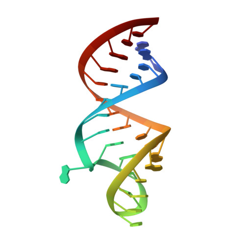
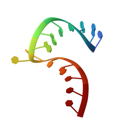
Identification and characterization of anion binding sites in RNA.
Kieft, J.S., Chase, E., Costantino, D.A., Golden, B.L.(2010) RNA 16: 1118-1123
- PubMed: 20410239
- DOI: https://doi.org/10.1261/rna.2072710
- Primary Citation of Related Structures:
3MJ3, 3MJA, 3MJB - PubMed Abstract:
Although RNA molecules are highly negatively charged, anions have been observed bound to RNA in crystal structures. It has been proposed that anion binding sites found within isolated RNAs represent regions of the molecule that could be involved in intermolecular interactions, indicating potential contact points for negatively charged amino acids from proteins or phosphate groups from an RNA. Several types of anion binding sites have been cataloged based on available structures. However, currently there is no method for unambiguously assigning anions to crystallographic electron density, and this has precluded more detailed analysis of RNA-anion interaction motifs and their significance. We therefore soaked selenate into two different types of RNA crystals and used the anomalous signal from these anions to identify binding sites in these RNA molecules unambiguously. Examination of these sites and comparison with other suspected anion binding sites reveals features of anion binding motifs, and shows that selenate may be a useful tool for studying RNA-anion interactions.
Organizational Affiliation:
Howard Hughes Medical Institute and Department of Biochemistry and Molecular Genetics, University of Colorado School of Medicine, Aurora, Colorado, 80045, USA. Jeffrey.Kieft@ucdenver.edu


