3M0L

Crystal structure of Pseudomonas stutzeri L-rhamnose isomerase mutant S329F in complex with D-psicose


Experimental Data Snapshot

  • Method: X-RAY DIFFRACTION
  • Resolution: 1.85 Å
  • R-Value Free: 0.210 
  • R-Value Work: 0.178 
  • R-Value Observed: 0.178 

Starting Model: experimental
View more details

wwPDB Validation   3D Report Full Report


Ligand Structure Quality Assessment 


This is version 1.3 of the entry. See complete history


Literature

Elucidation of the role of Ser329 and the C-terminal region in the catalytic activity of Pseudomonas stutzeri L-rhamnose isomerase

Yoshida, H.Takeda, K.Izumori, K.Kamitori, S.

(2010) Protein Eng Des Sel 23: 919-927

  • DOI: https://doi.org/10.1093/protein/gzq077
  • Primary Citation of Related Structures:  
    3M0H, 3M0L, 3M0M, 3M0V, 3M0X, 3M0Y

  • PubMed Abstract: 

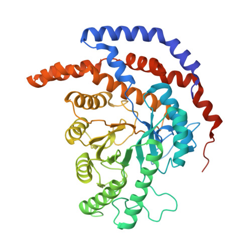
    Pseudomonas stutzeri l-rhamnose isomerase (l-RhI) is capable of catalyzing the isomerization between various aldoses and ketoses, showing high catalytic activity with broad substrate-specificity compared with Escherichia coli l-RhI. In a previous study, the crystal structure of P. stutzeri l-RhI revealed an active site comparable with that of E. coli l-RhI and d-xylose isomerases (d-XIs) with structurally conserved amino acids, but also with a different residue seemingly responsible for the specificity of P. stutzeri l-RhI, though the residue itself does not interact with the bound substrate. This residue, Ser329, corresponds to Phe336 in E. coli l-RhI and Lys294 in Actinoplanes missouriensis d-XI. To elucidate the role of Ser329 in P. stutzeri l-RhI, we constructed mutants, S329F (E. coli l-RhI type), S329K (A. missouriensis d-XI type), S329L and S329A. Analyses of the catalytic activity and crystal structure of the mutants revealed a hydroxyl group of Ser329 to be crucial for catalytic activity via interaction with a water molecule. In addition, in complexes with substrate, the mutants S329F and S329L exhibited significant electron density in the C-terminal region not observed in the wild-type P. stutzeri l-RhI. The C-terminal region of P. stutzeri l-RhI has flexibility and shows a flip-flop movement at the inter-molecular surface of the dimeric form.


  • Organizational Affiliation

    Division of Structural Biology, Life Science Research Center and Faculty of Medicine, Kagawa University, 1750-1 Ikenobe, Miki-cho, Kita-gun, Kagawa 761-0793, Japan.


Macromolecules
Find similar proteins by:  (by identity cutoff)  |  3D Structure
Entity ID: 1
MoleculeChains Sequence LengthOrganismDetailsImage
L-rhamnose isomerase
A, B, C, D
438Stutzerimonas stutzeriMutation(s): 2 
EC: 5.3.1.14
UniProt
Find proteins for Q75WH8 (Stutzerimonas stutzeri)
Explore Q75WH8 
Go to UniProtKB:  Q75WH8
Entity Groups  
Sequence Clusters30% Identity50% Identity70% Identity90% Identity95% Identity100% Identity
UniProt GroupQ75WH8
Sequence Annotations
Expand
  • Reference Sequence
Small Molecules
Ligands 2 Unique
IDChains Name / Formula / InChI Key2D Diagram3D Interactions
PSJ
Query on PSJ

Download Ideal Coordinates CCD File 
G [auth A],
J [auth B],
M [auth C],
P [auth D]
D-psicose
C6 H12 O6
BJHIKXHVCXFQLS-PUFIMZNGSA-N
MN
Query on MN

Download Ideal Coordinates CCD File 
E [auth A]
F [auth A]
H [auth B]
I [auth B]
K [auth C]
E [auth A],
F [auth A],
H [auth B],
I [auth B],
K [auth C],
L [auth C],
N [auth D],
O [auth D]
MANGANESE (II) ION
Mn
WAEMQWOKJMHJLA-UHFFFAOYSA-N
Experimental Data & Validation

Experimental Data

  • Method: X-RAY DIFFRACTION
  • Resolution: 1.85 Å
  • R-Value Free: 0.210 
  • R-Value Work: 0.178 
  • R-Value Observed: 0.178 
  • Space Group: P 1 21 1
Unit Cell:
Length ( Å )Angle ( ˚ )
a = 74.576α = 90
b = 104.117β = 106.21
c = 111.23γ = 90
Software Package:
Software NamePurpose
ADSCdata collection
MOLREPphasing
CNSrefinement
HKL-2000data reduction
HKL-2000data scaling

Structure Validation

View Full Validation Report


Ligand Structure Quality Assessment 


Entry History 

Deposition Data

Revision History  (Full details and data files)

  • Version 1.0: 2010-11-10
    Type: Initial release
  • Version 1.1: 2011-07-13
    Changes: Version format compliance
  • Version 1.2: 2021-11-10
    Changes: Database references, Derived calculations
  • Version 1.3: 2023-11-01
    Changes: Data collection, Refinement description