Crystal structures of truncated alphaA and alphaB crystallins reveal structural mechanisms of polydispersity important for eye lens function.
Laganowsky, A., Benesch, J.L., Landau, M., Ding, L., Sawaya, M.R., Cascio, D., Huang, Q., Robinson, C.V., Horwitz, J., Eisenberg, D.(2010) Protein Sci 19: 1031-1043
- PubMed: 20440841
- DOI: https://doi.org/10.1002/pro.380
- Primary Citation of Related Structures:
3L1E, 3L1F, 3L1G - PubMed Abstract:
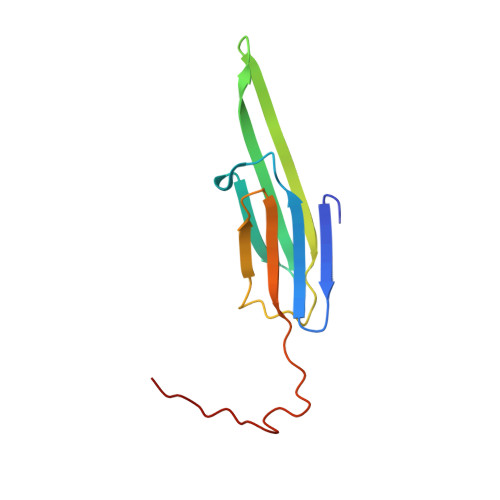
Small heat shock proteins alphaA and alphaB crystallin form highly polydisperse oligomers that frustrate protein aggregation, crystallization, and amyloid formation. Here, we present the crystal structures of truncated forms of bovine alphaA crystallin (AAC(59-163)) and human alphaB crystallin (ABC(68-162)), both containing the C-terminal extension that functions in chaperone action and oligomeric assembly. In both structures, the C-terminal extensions swap into neighboring molecules, creating runaway domain swaps. This interface, termed DS, enables crystallin polydispersity because the C-terminal extension is palindromic and thereby allows the formation of equivalent residue interactions in both directions. That is, we observe that the extension binds in opposite directions at the DS interfaces of AAC(59-163) and ABC(68-162). A second dimeric interface, termed AP, also enables polydispersity by forming an antiparallel beta sheet with three distinct registration shifts. These two polymorphic interfaces enforce polydispersity of alpha crystallin. This evolved polydispersity suggests molecular mechanisms for chaperone action and for prevention of crystallization, both necessary for transparency of eye lenses.
Organizational Affiliation:
Howard Hughes Medical Institute, UCLA-DOE Institute for Genomics and Proteomics, Los Angeles, California, USA.