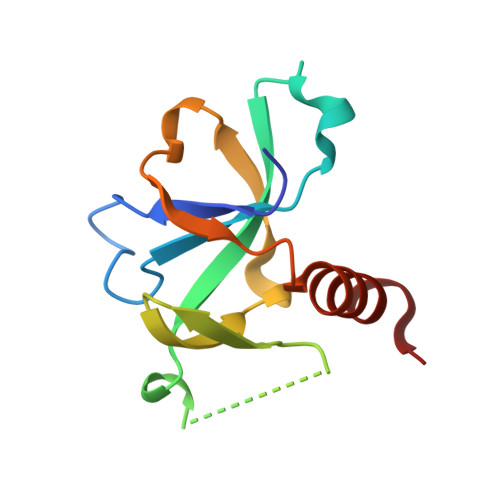
3JRZ

CcdBVfi-FormII-pH5.6


Experimental Data Snapshot

  • Method: X-RAY DIFFRACTION
  • Resolution: 1.70 Å
  • R-Value Free: 0.199 
  • R-Value Work: 0.168 
  • R-Value Observed: 0.169 

Starting Model: experimental
View more details

wwPDB Validation   3D Report Full Report


This is version 1.2 of the entry. See complete history


Literature

Structural and thermodynamic characterization of vibrio fischeri CCDB

De Jonge, N.Hohlweg, W.Garcia-Pino, A.Respondek, M.Buts, L.Haesaerts, S.Lah, J.Zangger, K.Loris, R.

(2010) J Biol Chem 285: 5606-5613

  • DOI: https://doi.org/10.1074/jbc.M109.068429
  • Primary Citation of Related Structures:  
    2KMT, 3JRZ, 3JSC

  • PubMed Abstract: 

    CcdB(Vfi) from Vibrio fischeri is a member of the CcdB family of toxins that poison covalent gyrase-DNA complexes. In solution CcdB(Vfi) is a dimer that unfolds to the corresponding monomeric components in a two-state fashion. In the unfolded state, the monomer retains a partial secondary structure. This observation correlates well with the crystal and NMR structures of the protein, which show a dimer with a hydrophobic core crossing the dimer interface. In contrast to its F plasmid homologue, CcdB(Vfi) possesses a rigid dimer interface, and the apparent relative rotations of the two subunits are due to structural plasticity of the monomer. CcdB(Vfi) shows a number of non-conservative substitutions compared with the F plasmid protein in both the CcdA and the gyrase binding sites. Although variation in the CcdA interaction site likely determines toxin-antitoxin specificity, substitutions in the gyrase-interacting region may have more profound functional implications.


  • Organizational Affiliation

    Structural Biology Brussels, and Department of Molecular and Cellular Interactions, Vrije Universiteit Brussel, Pleinlaan 2, B-1050 Brussels, Belgium.


Macromolecules
Find similar proteins by:  (by identity cutoff)  |  3D Structure
Entity ID: 1
MoleculeChains Sequence LengthOrganismDetailsImage
CcdB105Aliivibrio fischeriMutation(s): 0 
Gene Names: ccdB
UniProt
Find proteins for Q84B82 (Aliivibrio fischeri)
Explore Q84B82 
Go to UniProtKB:  Q84B82
Entity Groups  
Sequence Clusters30% Identity50% Identity70% Identity90% Identity95% Identity100% Identity
UniProt GroupQ84B82
Sequence Annotations
Expand
  • Reference Sequence
Experimental Data & Validation

Experimental Data

  • Method: X-RAY DIFFRACTION
  • Resolution: 1.70 Å
  • R-Value Free: 0.199 
  • R-Value Work: 0.168 
  • R-Value Observed: 0.169 
  • Space Group: C 1 2 1
Unit Cell:
Length ( Å )Angle ( ˚ )
a = 58.506α = 90
b = 43.624β = 110.01
c = 37.489γ = 90
Software Package:
Software NamePurpose
MAR345dtbdata collection
PHASERphasing
PHENIXrefinement
DENZOdata reduction
SCALEPACKdata scaling

Structure Validation

View Full Validation Report


Entry History 

Deposition Data

Revision History  (Full details and data files)

  • Version 1.0: 2009-12-01
    Type: Initial release
  • Version 1.1: 2011-07-13
    Changes: Version format compliance
  • Version 1.2: 2023-11-01
    Changes: Data collection, Database references, Refinement description