3J1T

High affinity dynein microtubule binding domain - tubulin complex


Experimental Data Snapshot

  • Method: ELECTRON MICROSCOPY
  • Resolution: 9.70 Å
  • Aggregation State: FILAMENT 
  • Reconstruction Method: HELICAL 

Starting Models: experimental
View more details

wwPDB Validation   3D Report Full Report


This is version 1.4 of the entry. See complete history


Literature

Structural basis for microtubule binding and release by dynein.

Redwine, W.B.Hernandez-Lopez, R.Zou, S.Huang, J.Reck-Peterson, S.L.Leschziner, A.E.

(2012) Science 337: 1532-1536

  • DOI: https://doi.org/10.1126/science.1224151
  • Primary Citation of Related Structures:  
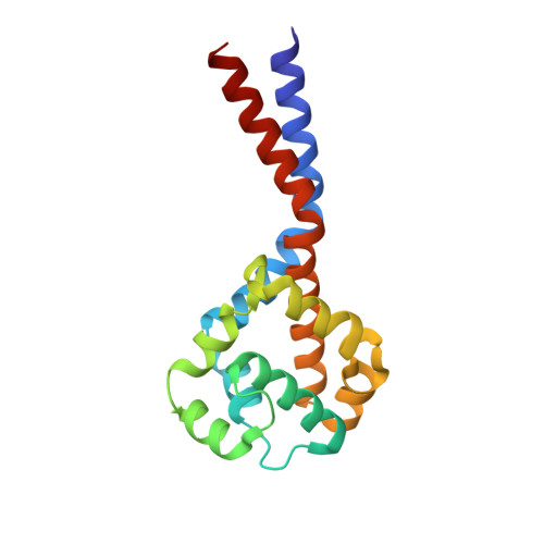
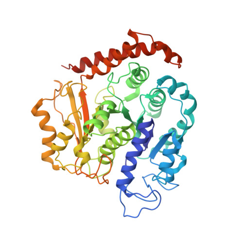
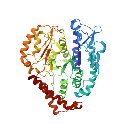
    3J1T, 3J1U

  • PubMed Abstract: 

    Cytoplasmic dynein is a microtubule-based motor required for intracellular transport and cell division. Its movement involves coupling cycles of track binding and release with cycles of force-generating nucleotide hydrolysis. How this is accomplished given the ~25 nanometers separating dynein's track- and nucleotide-binding sites is not understood. Here, we present a subnanometer-resolution structure of dynein's microtubule-binding domain bound to microtubules by cryo-electron microscopy that was used to generate a pseudo-atomic model of the complex with molecular dynamics. We identified large rearrangements triggered by track binding and specific interactions, confirmed by mutagenesis and single-molecule motility assays, which tune dynein's affinity for microtubules. Our results provide a molecular model for how dynein's binding to microtubules is communicated to the rest of the motor.


  • Organizational Affiliation

    Department of Molecular and Cellular Biology, Harvard University, 52 Oxford Street, Cambridge, MA 02138, United States.


Macromolecules
Find similar proteins by:  (by identity cutoff)  |  3D Structure
Entity ID: 1
MoleculeChains Sequence LengthOrganismDetailsImage
Cytoplasmic dynein 1 heavy chain 1, seryl t-RNA synthetase chimera164Mus musculusMutation(s): 0 
Gene Names: Dync1h1Dhc1Dnch1Dnchc1Dyhc
UniProt & NIH Common Fund Data Resources
Find proteins for Q9JHU4 (Mus musculus)
Explore Q9JHU4 
Go to UniProtKB:  Q9JHU4
IMPC:  MGI:103147
Entity Groups  
Sequence Clusters30% Identity50% Identity70% Identity90% Identity95% Identity100% Identity
UniProt GroupQ9JHU4
Sequence Annotations
Expand
  • Reference Sequence
Find similar proteins by:  (by identity cutoff)  |  3D Structure
Entity ID: 2
MoleculeChains Sequence LengthOrganismDetailsImage
Tubulin alpha-1B chain451Bos taurusMutation(s): 0 
UniProt
Find proteins for P81947 (Bos taurus)
Explore P81947 
Go to UniProtKB:  P81947
Entity Groups  
Sequence Clusters30% Identity50% Identity70% Identity90% Identity95% Identity100% Identity
UniProt GroupP81947
Sequence Annotations
Expand
  • Reference Sequence
Find similar proteins by:  (by identity cutoff)  |  3D Structure
Entity ID: 3
MoleculeChains Sequence LengthOrganismDetailsImage
Tubulin beta-2B chain427Bos taurusMutation(s): 0 
UniProt
Find proteins for Q6B856 (Bos taurus)
Explore Q6B856 
Go to UniProtKB:  Q6B856
Entity Groups  
Sequence Clusters30% Identity50% Identity70% Identity90% Identity95% Identity100% Identity
UniProt GroupQ6B856
Sequence Annotations
Expand
  • Reference Sequence
Experimental Data & Validation

Experimental Data

  • Method: ELECTRON MICROSCOPY
  • Resolution: 9.70 Å
  • Aggregation State: FILAMENT 
  • Reconstruction Method: HELICAL 
EM Software:
TaskSoftware PackageVersion
RECONSTRUCTIONFREALIGN
RECONSTRUCTIONSPIDER

Structure Validation

View Full Validation Report


Entry History 

Deposition Data

Revision History  (Full details and data files)

  • Version 1.0: 2012-09-26
    Type: Initial release
  • Version 1.1: 2012-10-03
    Changes: Database references
  • Version 1.2: 2014-08-27
    Changes: Other
  • Version 1.3: 2018-07-18
    Changes: Author supporting evidence, Data collection
  • Version 1.4: 2024-02-21
    Changes: Data collection, Database references, Refinement description