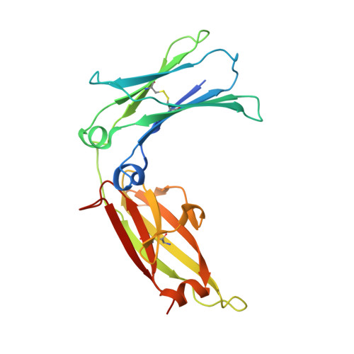
3HA0

Crystal structure of the IgE-Fc3-4 domains


Experimental Data Snapshot

  • Method: X-RAY DIFFRACTION
  • Resolution: 2.80 Å
  • R-Value Free: 0.280 
  • R-Value Work: 0.241 
  • R-Value Observed: 0.243 

Starting Model: experimental
View more details

wwPDB Validation   3D Report Full Report


This is version 2.3 of the entry. See complete history


Literature

Conformational flexibility in immunoglobulin E-Fc 3-4 revealed in multiple crystal forms.

Wurzburg, B.A.Jardetzky, T.S.

(2009) J Mol Biol 393: 176-190

  • DOI: https://doi.org/10.1016/j.jmb.2009.08.012
  • Primary Citation of Related Structures:  
    3H9Y, 3H9Z, 3HA0

  • PubMed Abstract: 

    The structure of immunoglobulin E (IgE)-Fc(3-4) has been solved in three new crystal forms, providing 13 snapshots of the Fc conformation and revealing a diverse range of open-closed motions among subunit chains and dimers. A more detailed analysis of the open-to-closed motion of IgE-Fc(3-4) was possible with so many structures, and the new structures allow a more thorough examination of the flexibility of IgE-Fc and its implications for receptor binding. The existence of a hydrophobic pocket at the elbow region of the Fc appears to be conformation dependent and suggests a means of regulating the IgE-Fc conformation (and potentially receptor binding) with small molecules.


  • Organizational Affiliation

    Department of Structural Biology, Stanford University, Stanford, CA 94305, USA.


Macromolecules
Find similar proteins by:  (by identity cutoff)  |  3D Structure
Entity ID: 1
MoleculeChains Sequence LengthOrganismDetailsImage
Ig epsilon chain C region
A, B, C, D, E
A, B, C, D, E, F
223Homo sapiensMutation(s): 2 
Gene Names: IGHE
UniProt & NIH Common Fund Data Resources
Find proteins for P01854 (Homo sapiens)
Explore P01854 
Go to UniProtKB:  P01854
PHAROS:  P01854
GTEx:  ENSG00000211891 
Entity Groups  
Sequence Clusters30% Identity50% Identity70% Identity90% Identity95% Identity100% Identity
UniProt GroupP01854
Glycosylation
Glycosylation Sites: 1Go to GlyGen: P01854-2
Sequence Annotations
Expand
  • Reference Sequence
Oligosaccharides

Help

Entity ID: 2
MoleculeChains Length2D Diagram Glycosylation3D Interactions
alpha-D-mannopyranose-(1-3)-[alpha-D-mannopyranose-(1-6)]beta-D-mannopyranose-(1-4)-2-acetamido-2-deoxy-beta-D-glucopyranose-(1-4)-2-acetamido-2-deoxy-beta-D-glucopyranose
G, H, I, J, K
G, H, I, J, K, L
5N-Glycosylation
Glycosylation Resources
GlyTouCan:  G22768VO
GlyCosmos:  G22768VO
GlyGen:  G22768VO
Experimental Data & Validation

Experimental Data

  • Method: X-RAY DIFFRACTION
  • Resolution: 2.80 Å
  • R-Value Free: 0.280 
  • R-Value Work: 0.241 
  • R-Value Observed: 0.243 
  • Space Group: P 1 21 1
Unit Cell:
Length ( Å )Angle ( ˚ )
a = 48.9α = 90
b = 104.9β = 96.2
c = 150γ = 90
Software Package:
Software NamePurpose
REFMACrefinement
SCALEPACKdata scaling
CNSrefinement
DENZOdata reduction
PDB_EXTRACTdata extraction
MAR345data collection
CNSphasing

Structure Validation

View Full Validation Report


Entry History 

Deposition Data

Revision History  (Full details and data files)

  • Version 1.0: 2009-09-08
    Type: Initial release
  • Version 1.1: 2011-07-13
    Changes: Advisory, Refinement description, Version format compliance
  • Version 1.2: 2017-11-01
    Changes: Refinement description
  • Version 1.3: 2019-07-24
    Changes: Data collection, Derived calculations, Refinement description
  • Version 2.0: 2020-07-29
    Type: Remediation
    Reason: Carbohydrate remediation
    Changes: Atomic model, Data collection, Database references, Derived calculations, Structure summary
  • Version 2.1: 2021-10-13
    Changes: Database references, Derived calculations, Structure summary
  • Version 2.2: 2023-09-06
    Changes: Data collection, Refinement description
  • Version 2.3: 2024-11-06
    Changes: Structure summary