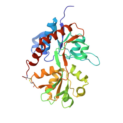
3H6U

Crystal structure of the iGluR2 ligand-binding core (S1S2J-N754S) in complex with glutamate and NS1493 at 1.85 A resolution


Experimental Data Snapshot

  • Method: X-RAY DIFFRACTION
  • Resolution: 1.85 Å
  • R-Value Free: 0.215 
  • R-Value Work: 0.181 
  • R-Value Observed: 0.181 

Starting Model: experimental
View more details

wwPDB Validation   3D Report Full Report


Ligand Structure Quality Assessment 


This is version 1.6 of the entry. See complete history


Literature

Distinct structural features of cyclothiazide are responsible for effects on peak current amplitude and desensitization kinetics at iGluR2.

Hald, H.Ahring, P.K.Timmermann, D.B.Liljefors, T.Gajhede, M.Kastrup, J.S.

(2009) J Mol Biol 391: 906-917

  • DOI: https://doi.org/10.1016/j.jmb.2009.07.002
  • Primary Citation of Related Structures:  
    3H6T, 3H6U, 3H6V, 3H6W

  • PubMed Abstract: 

    Ionotropic glutamate receptors (iGluRs) mediate fast excitatory neurotransmission. Upon glutamate application, 2-amino-3-(3-hydroxy-5-methyl-4-isoxazolyl)propionic acid receptors undergo rapid and almost complete desensitization that can be attenuated by positive allosteric modulators. The molecular mechanism of positive allosteric modulation has been elucidated previously by crystal structures of the ligand-binding core of iGluR2 in complex with, for example, cyclothiazide (CTZ). Here, we investigate the structure and function of CTZ and three closely related analogues NS1493, NS5206, and NS5217 at iGluR2, by X-ray crystallography and fast application patch-clamp electrophysiology. CTZ was the most efficacious and potent modulator of the four compounds on iGluR2(Q)(i) [E(max) normalized to response of glutamate: 754% (CTZ), 490% (NS1493), 399% (NS5206), and 476% (NS5217) and EC(50) in micromolar: 10 (CTZ), 26 (NS1493), 43 (NS5206), and 48 (NS5217)]. The four modulators divide into three groups according to efficacy and desensitization kinetics: (1) CTZ increases the peak current efficacy twice as much as the three analogues and nearly completely blocks receptor desensitization; (2) NS5206 and NS5217 have low efficacy and only attenuate desensitization partially; (3) NS1493 has low efficacy but nearly completely blocks receptor desensitization. A hydrophobic substituent at the 3-position of the 1,1-dioxo-3,4-dihydro-2H-benzo[e][1,2,4]thiadiazine ring system is important for compound efficacy, with the following ranking: norbornenyl (bicyclo[2.2.1]hept-2-ene)>cyclopentyl>methyl. The replacement of the norbornenyl moiety with a significantly less hydrophobic cyclopentane ring increases the flexibility of the modulator as the cyclopentane ring adopts various conformations at the iGluR2 allosteric binding site. The main structural feature responsible for a nearly complete block of desensitization is the presence of an NH hydrogen bond donor in the 4-position of the 1,1-dioxo-3,4-dihydro-2H-benzo[e][1,2,4]thiadiazine ring system, forming an anchoring hydrogen bond to Ser754. Therefore, the atom at the 4-position of CTZ seems to be a major determinant of receptor desensitization kinetics.


  • Organizational Affiliation

    Department of Medicinal Chemistry, University of Copenhagen, Denmark.


Macromolecules
Find similar proteins by:  (by identity cutoff)  |  3D Structure
Entity ID: 1
MoleculeChains Sequence LengthOrganismDetailsImage
Glutamate receptor 2263Rattus norvegicusMutation(s): 1 
Gene Names: Gria2Glur2
UniProt
Find proteins for P19491 (Rattus norvegicus)
Explore P19491 
Go to UniProtKB:  P19491
Entity Groups  
Sequence Clusters30% Identity50% Identity70% Identity90% Identity95% Identity100% Identity
UniProt GroupP19491
Sequence Annotations
Expand
  • Reference Sequence
Small Molecules
Ligands 5 Unique
IDChains Name / Formula / InChI Key2D Diagram3D Interactions
NS3
Query on NS3

Download Ideal Coordinates CCD File 
C [auth A](3S)-3-cyclopentyl-6-methyl-7-[(4-methylpiperazin-1-yl)sulfonyl]-3,4-dihydro-2H-1,2,4-benzothiadiazine 1,1-dioxide
C18 H28 N4 O4 S2
CUMKMTBOHBENJI-SFHVURJKSA-N
FLC
Query on FLC

Download Ideal Coordinates CCD File 
L [auth A]CITRATE ANION
C6 H5 O7
KRKNYBCHXYNGOX-UHFFFAOYSA-K
GLU
Query on GLU

Download Ideal Coordinates CCD File 
B [auth A]GLUTAMIC ACID
C5 H9 N O4
WHUUTDBJXJRKMK-VKHMYHEASA-N
SO4
Query on SO4

Download Ideal Coordinates CCD File 
D [auth A]
E [auth A]
F [auth A]
G [auth A]
H [auth A]
D [auth A],
E [auth A],
F [auth A],
G [auth A],
H [auth A],
I [auth A]
SULFATE ION
O4 S
QAOWNCQODCNURD-UHFFFAOYSA-L
GOL
Query on GOL

Download Ideal Coordinates CCD File 
J [auth A],
K [auth A]
GLYCEROL
C3 H8 O3
PEDCQBHIVMGVHV-UHFFFAOYSA-N
Experimental Data & Validation

Experimental Data

  • Method: X-RAY DIFFRACTION
  • Resolution: 1.85 Å
  • R-Value Free: 0.215 
  • R-Value Work: 0.181 
  • R-Value Observed: 0.181 
  • Space Group: P 21 21 2
Unit Cell:
Length ( Å )Angle ( ˚ )
a = 88.462α = 90
b = 64.086β = 90
c = 47.272γ = 90
Software Package:
Software NamePurpose
DENZOdata reduction
SCALEPACKdata scaling
CNSrefinement
PDB_EXTRACTdata extraction
REFMACphasing

Structure Validation

View Full Validation Report


Ligand Structure Quality Assessment 


Entry History 

Deposition Data

Revision History  (Full details and data files)

  • Version 1.0: 2009-07-28
    Type: Initial release
  • Version 1.1: 2011-07-13
    Changes: Non-polymer description, Version format compliance
  • Version 1.2: 2017-08-02
    Changes: Source and taxonomy
  • Version 1.3: 2017-11-01
    Changes: Refinement description
  • Version 1.4: 2021-10-13
    Changes: Data collection, Database references, Derived calculations
  • Version 1.5: 2023-09-06
    Changes: Data collection, Refinement description
  • Version 1.6: 2024-10-16
    Changes: Structure summary