


Kinetic and Structural studies on atazanavir-specific I50L drug-resistant HIV-1 protease mutant
Prabu-Jeyabalan, M., King, N., Bandaranayake, R., Nalivaika, E., Schiffer, C.To be published.
Experimental Data Snapshot
Starting Model: experimental
View more details
Entity ID: 1 | |||||
|---|---|---|---|---|---|
| Molecule | Chains | Sequence Length | Organism | Details | Image |
| Protease | A, B, C [auth U], D [auth V] | 99 | HIV-1 M:B_ARV2/SF2 | Mutation(s): 3 Gene Names: gag-pol, HIV-1 subtype B EC: 3.4.23.16 | 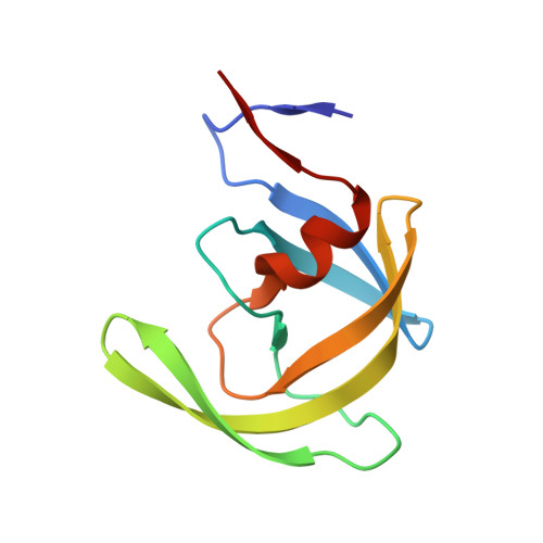 |
UniProt | |||||
Find proteins for P03369 (Human immunodeficiency virus type 1 group M subtype B (isolate ARV2/SF2)) Explore P03369 Go to UniProtKB: P03369 | |||||
Entity Groups | |||||
| Sequence Clusters | 30% Identity50% Identity70% Identity90% Identity95% Identity100% Identity | ||||
| UniProt Group | P03369 | ||||
Sequence AnnotationsExpand | |||||
| |||||
| Ligands 2 Unique | |||||
|---|---|---|---|---|---|
| ID | Chains | Name / Formula / InChI Key | 2D Diagram | 3D Interactions | |
| DR7 Query on DR7 | E [auth A], K [auth V] | (3S,8S,9S,12S)-3,12-BIS(1,1-DIMETHYLETHYL)-8-HYDROXY-4,11-DIOXO-9-(PHENYLMETHYL)-6-[[4-(2-PYRIDINYL)PHENYL]METHYL]-2,5,
6,10,13-PENTAAZATETRADECANEDIOIC ACID DIMETHYL ESTER C38 H52 N6 O7 AXRYRYVKAWYZBR-GASGPIRDSA-N |  | ||
| PO4 Query on PO4 | F [auth A] G [auth B] H [auth B] I [auth B] J [auth U] | PHOSPHATE ION O4 P NBIIXXVUZAFLBC-UHFFFAOYSA-K |  | ||
| Length ( Å ) | Angle ( ˚ ) |
|---|---|
| a = 51.231 | α = 90 |
| b = 59.54 | β = 82.22 |
| c = 59.863 | γ = 90 |
| Software Name | Purpose |
|---|---|
| DENZO | data reduction |
| SCALEPACK | data scaling |
| REFMAC | refinement |
| PDB_EXTRACT | data extraction |
| AMoRE | phasing |
RCSB PDB (citation) is hosted by
RCSB PDB is a member of the

