3DOR

Crystal Structure of mature CPAF


Experimental Data Snapshot

  • Method: X-RAY DIFFRACTION
  • Resolution: 2.20 Å
  • R-Value Free: 0.262 
  • R-Value Work: 0.228 
  • R-Value Observed: 0.228 

Starting Model: experimental
View more details

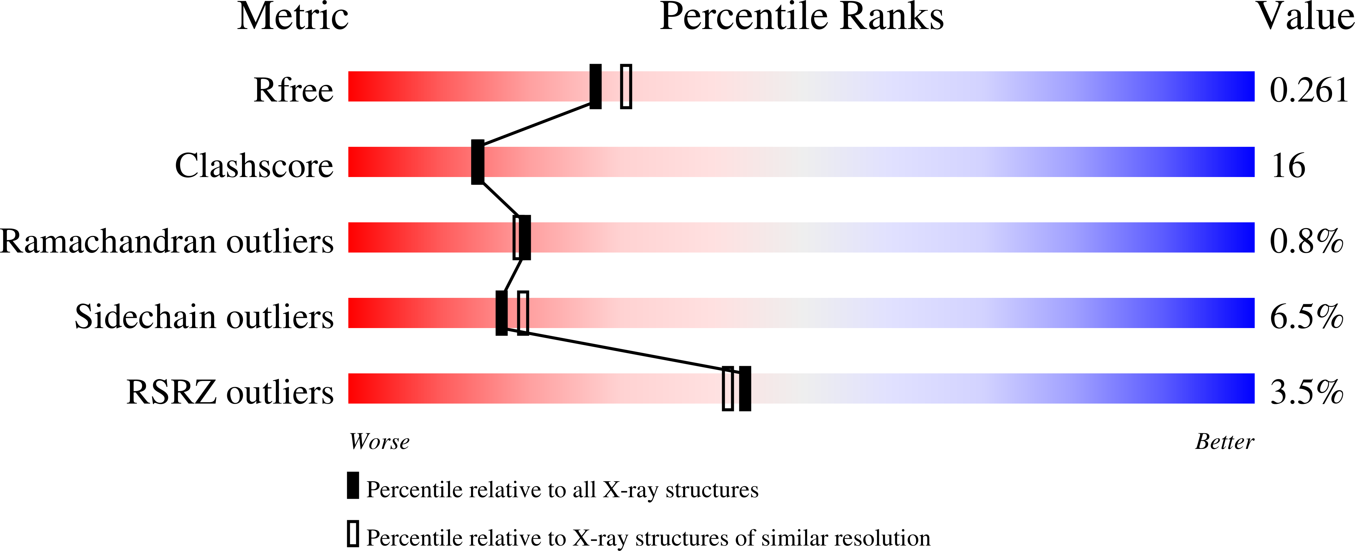
wwPDB Validation   3D Report Full Report


This is version 1.2 of the entry. See complete history


Literature

Structural basis for activation and inhibition of the secreted chlamydia protease CPAF

Huang, Z.Feng, Y.Chen, D.Wu, X.Huang, S.Wang, X.Xiao, X.Li, W.Huang, N.Gu, L.Zhong, G.Chai, J.

(2008) Cell Host Microbe 4: 529-542

  • DOI: https://doi.org/10.1016/j.chom.2008.10.005
  • Primary Citation of Related Structures:  
    3DJA, 3DOR, 3DPM, 3DPN

  • PubMed Abstract: 

    The obligate intracellular pathogen Chlamydia trachomatis is the most common cause of sexually transmitted bacterial disease. It secretes a protease known as chlamydial protease/proteasome-like activity factor (CPAF) that degrades many host molecules and plays a major role in Chlamydia pathogenesis. Here, we show that mature CPAF is a homodimer of the catalytic domains, each of which comprises two distinct subunits. Dormancy of the CPAF zymogen is maintained by an internal inhibitory segment that binds the CPAF active site and blocks its homodimerization. CPAF activation is initiated by trans-autocatalytic cleavage, which induces homodimerization and conformational changes that assemble the catalytic triad. This assembly leads to two autocatalytic cleavages and removal of the inhibitory segment, enabling full CPAF activity. CPAF is covalently bound and inhibited by the proteasome inhibitor lactacystin. These results reveal the activation mechanism of the CPAF serine protease and suggest new opportunities for anti-Chlamydia drug development.


  • Organizational Affiliation

    College of Biological Sciences, China Agricultural University, Beijing, China.


Macromolecules
Find similar proteins by:  (by identity cutoff)  |  3D Structure
Entity ID: 1
MoleculeChains Sequence LengthOrganismDetailsImage
Protein CT_858
A, B
583Chlamydia trachomatisMutation(s): 0 
UniProt
Find proteins for O84866 (Chlamydia trachomatis serovar D (strain ATCC VR-885 / DSM 19411 / UW-3/Cx))
Explore O84866 
Go to UniProtKB:  O84866
Entity Groups  
Sequence Clusters30% Identity50% Identity70% Identity90% Identity95% Identity100% Identity
UniProt GroupO84866
Sequence Annotations
Expand
  • Reference Sequence
Small Molecules
Ligands 1 Unique
IDChains Name / Formula / InChI Key2D Diagram3D Interactions
SO4
Query on SO4

Download Ideal Coordinates CCD File 
C [auth A]
D [auth A]
E [auth A]
F [auth A]
G [auth A]
C [auth A],
D [auth A],
E [auth A],
F [auth A],
G [auth A],
H [auth A],
I [auth A],
J [auth A],
K [auth A],
L [auth B],
M [auth B],
N [auth B],
O [auth B],
P [auth B],
Q [auth B]
SULFATE ION
O4 S
QAOWNCQODCNURD-UHFFFAOYSA-L
Experimental Data & Validation

Experimental Data

  • Method: X-RAY DIFFRACTION
  • Resolution: 2.20 Å
  • R-Value Free: 0.262 
  • R-Value Work: 0.228 
  • R-Value Observed: 0.228 
  • Space Group: P 21 21 21
Unit Cell:
Length ( Å )Angle ( ˚ )
a = 61.3α = 90
b = 152.37β = 90
c = 162.58γ = 90
Software Package:
Software NamePurpose
HKL-2000data collection
SOLVEphasing
CNSrefinement
HKL-2000data reduction
HKL-2000data scaling

Structure Validation

View Full Validation Report


Entry History 

Deposition Data

Revision History  (Full details and data files)

  • Version 1.0: 2009-01-13
    Type: Initial release
  • Version 1.1: 2011-07-13
    Changes: Version format compliance
  • Version 1.2: 2023-11-01
    Changes: Data collection, Database references, Derived calculations, Refinement description