3C4H

Human poly(ADP-ribose) polymerase 3, catalytic fragment in complex with an inhibitor DR2313


Experimental Data Snapshot

  • Method: X-RAY DIFFRACTION
  • Resolution: 2.10 Å
  • R-Value Free: 0.242 
  • R-Value Work: 0.174 
  • R-Value Observed: 0.178 

Starting Model: experimental
View more details

wwPDB Validation   3D Report Full Report


Ligand Structure Quality Assessment 


This is version 1.2 of the entry. See complete history


Literature

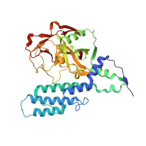
Structural basis for inhibitor specificity in human poly(ADP-ribose) polymerase-3.

Lehtio, L.Jemth, A.S.Collins, R.Loseva, O.Johansson, A.Markova, N.Hammarstrom, M.Flores, A.Holmberg-Schiavone, L.Weigelt, J.Helleday, T.Schuler, H.Karlberg, T.

(2009) J Med Chem 52: 3108-3111

  • DOI: https://doi.org/10.1021/jm900052j
  • Primary Citation of Related Structures:  
    3C49, 3C4H, 3CE0, 3FHB

  • PubMed Abstract: 

    Poly(ADP-ribose) polymerases (PARPs) activate DNA repair mechanisms upon stress- and cytotoxin-induced DNA damage, and inhibition of PARP activity is a lead in cancer drug therapy. We present a structural and functional analysis of the PARP domain of human PARP-3 in complex with several inhibitors. Of these, KU0058948 is the strongest inhibitor of PARP-3 activity. The presented crystal structures highlight key features for potent inhibitor binding and suggest routes for creating isoenzyme-specific PARP inhibitors.


  • Organizational Affiliation

    Structural Genomics Consortium, Department of Medical Biochemistry and Biophysics, Karolinska Institutet, SE-17177 Stockholm, Sweden.


Macromolecules
Find similar proteins by:  (by identity cutoff)  |  3D Structure
Entity ID: 1
MoleculeChains Sequence LengthOrganismDetailsImage
Poly(ADP-ribose) polymerase 3357Homo sapiensMutation(s): 0 
Gene Names: PARP3ADPRT3ADPRTL3
EC: 2.4.2.30
UniProt & NIH Common Fund Data Resources
Find proteins for Q9Y6F1 (Homo sapiens)
Explore Q9Y6F1 
Go to UniProtKB:  Q9Y6F1
PHAROS:  Q9Y6F1
GTEx:  ENSG00000041880 
Entity Groups  
Sequence Clusters30% Identity50% Identity70% Identity90% Identity95% Identity100% Identity
UniProt GroupQ9Y6F1
Sequence Annotations
Expand
  • Reference Sequence
Small Molecules
Ligands 2 Unique
IDChains Name / Formula / InChI Key2D Diagram3D Interactions
DRL
Query on DRL

Download Ideal Coordinates CCD File 
B [auth A]2-methyl-3,5,7,8-tetrahydro-4H-thiopyrano[4,3-d]pyrimidin-4-one
C8 H10 N2 O S
HRYKZAKEAVZGJD-UHFFFAOYSA-N
DMS
Query on DMS

Download Ideal Coordinates CCD File 
C [auth A]DIMETHYL SULFOXIDE
C2 H6 O S
IAZDPXIOMUYVGZ-UHFFFAOYSA-N
Binding Affinity Annotations 
IDSourceBinding Affinity
DRL PDBBind:  3C4H Kd: 2000 (nM) from 1 assay(s)
BindingDB:  3C4H Kd: 2000 (nM) from 1 assay(s)
Experimental Data & Validation

Experimental Data

  • Method: X-RAY DIFFRACTION
  • Resolution: 2.10 Å
  • R-Value Free: 0.242 
  • R-Value Work: 0.174 
  • R-Value Observed: 0.178 
  • Space Group: P 1 21 1
Unit Cell:
Length ( Å )Angle ( ˚ )
a = 54.91α = 90
b = 57.33β = 112.36
c = 56.26γ = 90
Software Package:
Software NamePurpose
REFMACrefinement
MxCuBEdata collection
XDSdata reduction
XSCALEdata scaling
MOLREPphasing

Structure Validation

View Full Validation Report


Ligand Structure Quality Assessment 


Entry History 

Revision History  (Full details and data files)

  • Version 1.0: 2008-02-12
    Type: Initial release
  • Version 1.1: 2011-07-13
    Changes: Advisory, Version format compliance
  • Version 1.2: 2023-08-30
    Changes: Data collection, Database references, Derived calculations, Refinement description