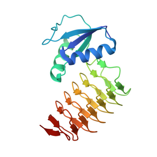
3BSY

PglD from Campylobacter jejuni, NCTC 11168, in complex with acetyl coenzyme A


Experimental Data Snapshot

  • Method: X-RAY DIFFRACTION
  • Resolution: 1.80 Å
  • R-Value Free: 0.220 
  • R-Value Work: 0.184 
  • R-Value Observed: 0.186 

Starting Model: experimental
View more details

wwPDB Validation   3D Report Full Report


Ligand Structure Quality Assessment 


This is version 1.4 of the entry. See complete history


Literature

Crystal structure and catalytic mechanism of PglD from Campylobacter jejuni.

Olivier, N.B.Imperiali, B.

(2008) J Biol Chem 283: 27937-27946

  • DOI: https://doi.org/10.1074/jbc.M801207200
  • Primary Citation of Related Structures:  
    3BSS, 3BSW, 3BSY

  • PubMed Abstract: 

    The carbohydrate 2, 4-diacetamido-2, 4, 6-trideoxy-alpha-D-glucopyranose (BacAc(2)) is found in a variety of eubacterial pathogens. In Campylobacter jejuni, PglD acetylates the C4 amino group on UDP-2-acetamido-4-amino-2, 4, 6-trideoxy-alpha-D-glucopyranose (UDP-4-amino-sugar) to form UDP-BacAc(2). Sequence analysis predicts PglD to be a member of the left-handed beta helix family of enzymes. However, poor sequence homology between PglD and left-handed beta helix enzymes with existing structural data precludes unambiguous identification of the active site. The co-crystal structures of PglD in the presence of citrate, acetyl coenzyme A, or the UDP-4-amino-sugar were solved. The biological assembly is a trimer with one active site formed between two protomers. Residues lining the active site were identified, and results from functional assays on alanine mutants suggest His-125 is critical for catalysis, whereas His-15 and His-134 are involved in substrate binding. These results are discussed in the context of implications for proteins homologous to PglD in other pathogens.


  • Organizational Affiliation

    Departments of Chemistry and Biology, Massachusetts Institute of Technology, Cambridge, Massachusetts 02139.


Macromolecules
Find similar proteins by:  (by identity cutoff)  |  3D Structure
Entity ID: 1
MoleculeChains Sequence LengthOrganismDetailsImage
Acetyltransferase
A, B, C
198Campylobacter jejuniMutation(s): 0 
Gene Names: pglD
EC: 2.7.7.23 (PDB Primary Data), 2.3.1.203 (UniProt)
UniProt
Find proteins for Q0P9D1 (Campylobacter jejuni subsp. jejuni serotype O:2 (strain ATCC 700819 / NCTC 11168))
Explore Q0P9D1 
Go to UniProtKB:  Q0P9D1
Entity Groups  
Sequence Clusters30% Identity50% Identity70% Identity90% Identity95% Identity100% Identity
UniProt GroupQ0P9D1
Sequence Annotations
Expand
  • Reference Sequence
Experimental Data & Validation

Experimental Data

  • Method: X-RAY DIFFRACTION
  • Resolution: 1.80 Å
  • R-Value Free: 0.220 
  • R-Value Work: 0.184 
  • R-Value Observed: 0.186 
  • Space Group: P 21 21 21
Unit Cell:
Length ( Å )Angle ( ˚ )
a = 55.871α = 90
b = 91.862β = 90
c = 125.195γ = 90
Software Package:
Software NamePurpose
DENZOdata reduction
SCALEPACKdata scaling
PHASERphasing
DMphasing
REFMACrefinement
PDB_EXTRACTdata extraction
HKL-2000data collection
HKL-2000data reduction
HKL-2000data scaling

Structure Validation

View Full Validation Report


Ligand Structure Quality Assessment 


Entry History 

Deposition Data

Revision History  (Full details and data files)

  • Version 1.0: 2008-07-29
    Type: Initial release
  • Version 1.1: 2011-07-13
    Changes: Version format compliance
  • Version 1.2: 2014-11-12
    Changes: Structure summary
  • Version 1.3: 2017-10-25
    Changes: Refinement description
  • Version 1.4: 2023-08-30
    Changes: Data collection, Database references, Derived calculations, Refinement description