3BL6

Crystal structure of Staphylococcus aureus 5'-methylthioadenosine/S-adenosylhomocysteine nucleosidase in complex with formycin A


Experimental Data Snapshot

  • Method: X-RAY DIFFRACTION
  • Resolution: 1.70 Å
  • R-Value Free: 0.187 
  • R-Value Work: 0.181 

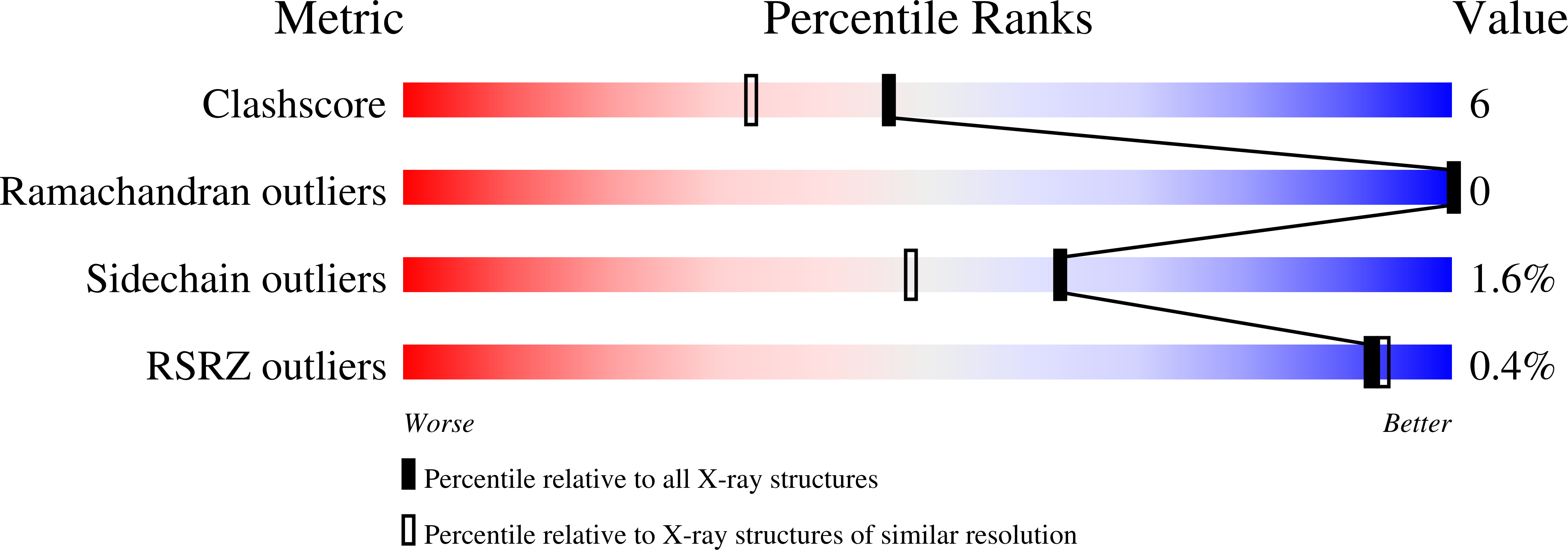
wwPDB Validation   3D Report Full Report


Ligand Structure Quality Assessment 


This is version 1.2 of the entry. See complete history


Literature

Structure of Staphylococcus aureus 5'-methylthioadenosine/S-adenosylhomocysteine nucleosidase

Siu, K.K.Lee, J.E.Smith, G.D.Horvatin-Mrakovcic, C.Howell, P.L.

(2008) Acta Crystallogr Sect F Struct Biol Cryst Commun 64: 343-350

  • DOI: https://doi.org/10.1107/S1744309108009275
  • Primary Citation of Related Structures:  
    3BL6, 3DF9

  • PubMed Abstract: 

    5'-Methylthioadenosine/S-adenosylhomocysteine nucleosidase (MTAN) catalyzes the irreversible cleavage of the glycosidic bond in 5'-methylthioadenosine (MTA) and S-adenosylhomocysteine (SAH) and plays a key role in four metabolic processes: biological methylation, polyamine biosynthesis, methionine recycling and bacterial quorum sensing. The absence of the nucleosidase in mammalian species has implicated this enzyme as a target for antimicrobial drug design. MTAN from the pathogenic bacterium Staphylococcus aureus (SaMTAN) has been kinetically characterized and its structure has been determined in complex with the transition-state analogue formycin A (FMA) at 1.7 A resolution. A comparison of the SaMTAN-FMA complex with available Escherichia coli MTAN structures shows strong conservation of the overall structure and in particular of the active site. The presence of an extra water molecule, which forms a hydrogen bond to the O4' atom of formycin A in the active site of SaMTAN, produces electron withdrawal from the ribosyl group and may explain the lower catalytic efficiency that SaMTAN exhibits when metabolizing MTA and SAH relative to the E. coli enzyme. The implications of this structure for broad-based antibiotic design are discussed.


  • Organizational Affiliation

    Program in Molecular Structure and Function, Research Institute, The Hospital for Sick Children, 555 University Avenue, Toronto, Ontario M5G 1X8, Canada.


Macromolecules
Find similar proteins by:  (by identity cutoff)  |  3D Structure
Entity ID: 1
MoleculeChains Sequence LengthOrganismDetailsImage
5'-methylthioadenosine nucleosidase/S-adenosylhomocysteine nucleosidase230Staphylococcus aureusMutation(s): 0 
Gene Names: pfs
EC: 3.2.2.9 (PDB Primary Data), 3.2.2.16 (PDB Primary Data)
UniProt
Find proteins for Q99TQ0 (Staphylococcus aureus (strain Mu50 / ATCC 700699))
Explore Q99TQ0 
Go to UniProtKB:  Q99TQ0
Entity Groups  
Sequence Clusters30% Identity50% Identity70% Identity90% Identity95% Identity100% Identity
UniProt GroupQ99TQ0
Sequence Annotations
Expand
  • Reference Sequence
Small Molecules
Ligands 1 Unique
IDChains Name / Formula / InChI Key2D Diagram3D Interactions
FMC
Query on FMC

Download Ideal Coordinates CCD File 
B [auth A](1S)-1-(7-amino-1H-pyrazolo[4,3-d]pyrimidin-3-yl)-1,4-anhydro-D-ribitol
C10 H13 N5 O4
KBHMEHLJSZMEMI-KSYZLYKTSA-N
Experimental Data & Validation

Experimental Data

  • Method: X-RAY DIFFRACTION
  • Resolution: 1.70 Å
  • R-Value Free: 0.187 
  • R-Value Work: 0.181 
  • Space Group: P 21 21 2
Unit Cell:
Length ( Å )Angle ( ˚ )
a = 58.25α = 90
b = 81.67β = 90
c = 45.46γ = 90
Software Package:
Software NamePurpose
d*TREKdata scaling
CNSrefinement
PDB_EXTRACTdata extraction
CrystalCleardata collection
d*TREKdata reduction
CNSphasing

Structure Validation

View Full Validation Report


Ligand Structure Quality Assessment 


Entry History 

Deposition Data

Revision History  (Full details and data files)

  • Version 1.0: 2008-06-10
    Type: Initial release
  • Version 1.1: 2011-07-13
    Changes: Version format compliance
  • Version 1.2: 2024-02-21
    Changes: Data collection, Database references, Derived calculations