3AJE

Crystal structure of S. tokodaii Sua5 complexed with L-threonine and AMPPNP


Experimental Data Snapshot

  • Method: X-RAY DIFFRACTION
  • Resolution: 1.80 Å
  • R-Value Free: 0.238 
  • R-Value Work: 0.198 
  • R-Value Observed: 0.198 

Starting Model: experimental
View more details

wwPDB Validation   3D Report Full Report


Ligand Structure Quality Assessment 


This is version 1.3 of the entry. See complete history


Literature

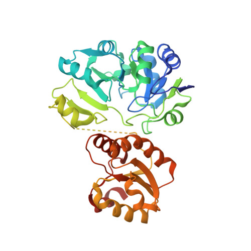
Crystal structure of Sulfolobus tokodaii Sua5 complexed with L-threonine and AMPPNP

Kuratani, M.Kasai, T.Akasaka, R.Higashijima, K.Terada, T.Kigawa, T.Shinkai, A.Bessho, Y.Yokoyama, S.

(2011) Proteins 79: 2065-2075

  • DOI: https://doi.org/10.1002/prot.23026
  • Primary Citation of Related Structures:  
    3AJE

  • PubMed Abstract: 

    The hypermodified nucleoside N(6)-threonylcarbamoyladenosine resides at position 37 of tRNA molecules bearing U at position 36 and maintains translational fidelity in the three kingdoms of life. The N(6)-threonylcarbamoyl moiety is composed of L-threonine and bicarbonate, and its synthesis was genetically shown to require YrdC/Sua5. YrdC/Sua5 binds to tRNA and ATP. In this study, we analyzed the L-threonine-binding mode of Sua5 from the archaeon Sulfolobus tokodaii. Isothermal titration calorimetry measurements revealed that S. tokodaii Sua5 binds L-threonine more strongly than L-serine and glycine. The Kd values of Sua5 for L-threonine and L-serine are 9.3 μM and 2.6 mM, respectively. We determined the crystal structure of S. tokodaii Sua5, complexed with AMPPNP and L-threonine, at 1.8 Å resolution. The L-threonine is bound next to AMPPNP in the same pocket of the N-terminal domain. Thr118 and two water molecules form hydrogen bonds with AMPPNP in a unique manner for adenine-specific recognition. The carboxyl group and the side-chain hydroxyl and methyl groups of L-threonine are buried deep in the pocket, whereas the amino group faces AMPPNP. The L-threonine is located in a suitable position to react together with ATP for the synthesis of N(6)-threonylcarbamoyladenosine.


  • Organizational Affiliation

    RIKEN Systems and Structural Biology Center, Yokohama 230-0045, Japan.


Macromolecules
Find similar proteins by:  (by identity cutoff)  |  3D Structure
Entity ID: 1
MoleculeChains Sequence LengthOrganismDetailsImage
Putative uncharacterized protein ST1526352Sulfurisphaera tokodaii str. 7Mutation(s): 0 
Gene Names: ST1526
UniProt
Find proteins for Q970S6 (Sulfurisphaera tokodaii (strain DSM 16993 / JCM 10545 / NBRC 100140 / 7))
Explore Q970S6 
Go to UniProtKB:  Q970S6
Entity Groups  
Sequence Clusters30% Identity50% Identity70% Identity90% Identity95% Identity100% Identity
UniProt GroupQ970S6
Sequence Annotations
Expand
  • Reference Sequence
Small Molecules
Ligands 3 Unique
IDChains Name / Formula / InChI Key2D Diagram3D Interactions
ANP
Query on ANP

Download Ideal Coordinates CCD File 
C [auth A]PHOSPHOAMINOPHOSPHONIC ACID-ADENYLATE ESTER
C10 H17 N6 O12 P3
PVKSNHVPLWYQGJ-KQYNXXCUSA-N
THR
Query on THR

Download Ideal Coordinates CCD File 
B [auth A]THREONINE
C4 H9 N O3
AYFVYJQAPQTCCC-GBXIJSLDSA-N
NO3
Query on NO3

Download Ideal Coordinates CCD File 
D [auth A]
E [auth A]
F [auth A]
G [auth A]
H [auth A]
D [auth A],
E [auth A],
F [auth A],
G [auth A],
H [auth A],
I [auth A]
NITRATE ION
N O3
NHNBFGGVMKEFGY-UHFFFAOYSA-N
Binding Affinity Annotations 
IDSourceBinding Affinity
THR PDBBind:  3AJE Kd: 9300 (nM) from 1 assay(s)
Experimental Data & Validation

Experimental Data

  • Method: X-RAY DIFFRACTION
  • Resolution: 1.80 Å
  • R-Value Free: 0.238 
  • R-Value Work: 0.198 
  • R-Value Observed: 0.198 
  • Space Group: C 2 2 2
Unit Cell:
Length ( Å )Angle ( ˚ )
a = 85.35α = 90
b = 121β = 90
c = 68.01γ = 90
Software Package:
Software NamePurpose
HKL-2000data collection
MOLREPphasing
CNSrefinement
HKL-2000data reduction
SCALEPACKdata scaling

Structure Validation

View Full Validation Report


Ligand Structure Quality Assessment 


Entry History 

Deposition Data

Revision History  (Full details and data files)

  • Version 1.0: 2011-04-13
    Type: Initial release
  • Version 1.1: 2011-07-13
    Changes: Version format compliance
  • Version 1.2: 2014-01-29
    Changes: Database references
  • Version 1.3: 2023-11-01
    Changes: Data collection, Database references, Derived calculations, Refinement description