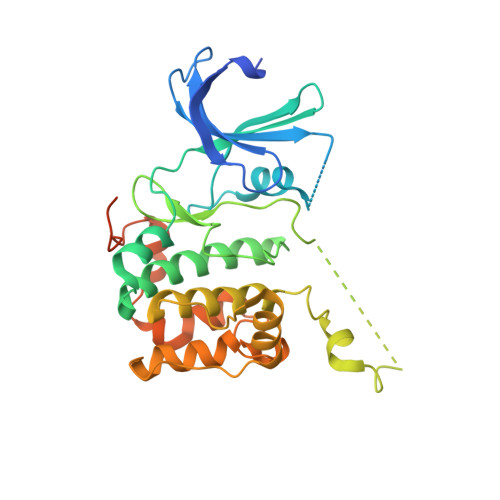
Structural basis of human p70 ribosomal S6 kinase-1 regulation by activation loop phosphorylation.
Sunami, T., Byrne, N., Diehl, R.E., Funabashi, K., Hall, D.L., Ikuta, M., Patel, S.B., Shipman, J.M., Smith, R.F., Takahashi, I., Zugay-Murphy, J., Iwasawa, Y., Lumb, K.J., Munshi, S.K., Sharma, S.(2010) J Biol Chem 285: 4587-4594
- PubMed: 19864428
- DOI: https://doi.org/10.1074/jbc.M109.040667
- Primary Citation of Related Structures:
3A60, 3A61, 3A62 - PubMed Abstract:
p70 ribosomal S6 kinase (p70S6K) is a downstream effector of the mTOR signaling pathway involved in cell proliferation, cell growth, cell-cycle progression, and glucose homeostasis. Multiple phosphorylation events within the catalytic, autoinhibitory, and hydrophobic motif domains contribute to the regulation of p70S6K. We report the crystal structures of the kinase domain of p70S6K1 bound to staurosporine in both the unphosphorylated state and in the 3'-phosphoinositide-dependent kinase-1-phosphorylated state in which Thr-252 of the activation loop is phosphorylated. Unphosphorylated p70S6K1 exists in two crystal forms, one in which the p70S6K1 kinase domain exists as a monomer and the other as a domain-swapped dimer. The crystal structure of the partially activated kinase domain that is phosphorylated within the activation loop reveals conformational ordering of the activation loop that is consistent with a role in activation. The structures offer insights into the structural basis of the 3'-phosphoinositide-dependent kinase-1-induced activation of p70S6K and provide a platform for the rational structure-guided design of specific p70S6K inhibitors.
Organizational Affiliation:
Department of Chemistry, Tsukuba Research Institute, Banyu Pharmaceutical Company, Limited, Tsukuba, Ibaraki, 300-2611, Japan. sunami.tomoko@jaea.go.jp


