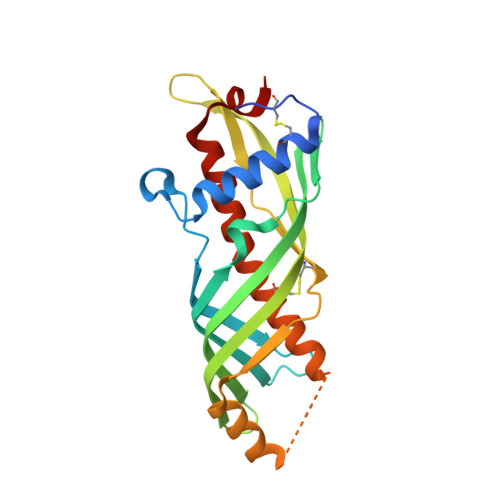
Crystal structure of silkworm Bombyx mori JHBP in complex with 2-methyl-2,4-pentanediol: plasticity of JH-binding pocket and ligand-induced conformational change of the second cavity in JHBP
Fujimoto, Z., Suzuki, R., Shiotsuki, T., Tsuchiya, W., Tase, A., Momma, M., Yamazaki, T.(2013) PLoS One 8: e56261-e56261
- PubMed: 23437107
- DOI: https://doi.org/10.1371/journal.pone.0056261
- Primary Citation of Related Structures:
3A1Z - PubMed Abstract:
Juvenile hormones (JHs) control a diversity of crucial life events in insects. In Lepidoptera which major agricultural pests belong to, JH signaling is critically controlled by a species-specific high-affinity, low molecular weight JH-binding protein (JHBP) in hemolymph, which transports JH from the site of its synthesis to target tissues. Hence, JHBP is expected to be an excellent target for the development of novel specific insect growth regulators (IGRs) and insecticides. A better understanding of the structural biology of JHBP should pave the way for the structure-based drug design of such compounds. Here, we report the crystal structure of the silkworm Bombyx mori JHBP in complex with two molecules of 2-methyl-2,4-pentanediol (MPD), one molecule (MPD1) bound in the JH-binding pocket while the other (MPD2) in a second cavity. Detailed comparison with the apo-JHBP and JHBP-JH II complex structures previously reported by us led to a number of intriguing findings. First, the JH-binding pocket changes its size in a ligand-dependent manner due to flexibility of the gate α1 helix. Second, MPD1 mimics interactions of the epoxide moiety of JH previously observed in the JHBP-JH complex, and MPD can compete with JH in binding to the JH-binding pocket. We also confirmed that methoprene, which has an MPD-like structure, inhibits the complex formation between JHBP and JH while the unepoxydated JH III (methyl farnesoate) does not. These findings may open the door to the development of novel IGRs targeted against JHBP. Third, binding of MPD to the second cavity of JHBP induces significant conformational changes accompanied with a cavity expansion. This finding, together with MPD2-JHBP interaction mechanism identified in the JHBP-MPD complex, should provide important guidance in the search for the natural ligand of the second cavity.
Organizational Affiliation:
Biomolecular Research Unit, National Institute of Agrobiological Sciences, Tsukuba, Ibaraki, Japan.