3ZOI

ISOPENICILLIN N SYNTHASE WITH AC-O-METHYL-D-THREONINE


Experimental Data Snapshot

  • Method: X-RAY DIFFRACTION
  • Resolution: 1.82 Å
  • R-Value Free: 0.202 
  • R-Value Work: 0.160 
  • R-Value Observed: 0.162 

Starting Model: experimental
View more details

wwPDB Validation   3D Report Full Report


Ligand Structure Quality Assessment 


This is version 1.4 of the entry. See complete history


Literature

The Crystal Structure of an Isopenicillin N Synthase Complex with an Ethereal Substrate Analogue Reveals Water in the Oxygen Binding Site.

Clifton, I.J.Ge, W.Adlington, R.M.Baldwin, J.E.Rutledge, P.J.

(2013) FEBS Lett 587: 2705

  • DOI: https://doi.org/10.1016/j.febslet.2013.07.016
  • Primary Citation of Related Structures:  
    3ZOI

  • PubMed Abstract: 

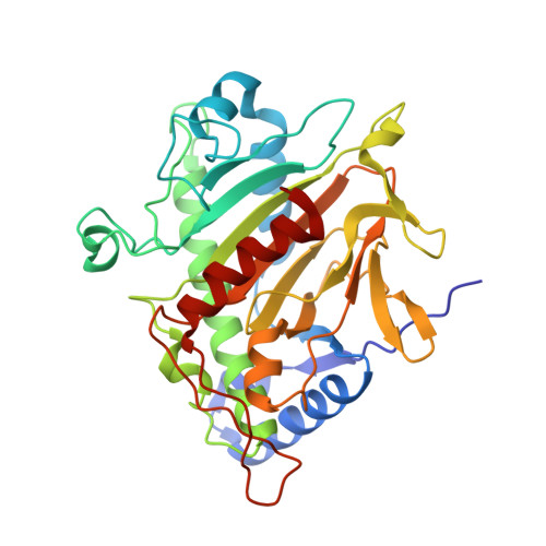
    Isopenicillin N synthase (IPNS) is a non-heme iron oxidase central to the biosynthesis of β-lactam antibiotics. IPNS converts the tripeptide δ-(L-α-aminoadipoyl)-L-cysteinyl-D-valine (ACV) to isopenicillin N while reducing molecular oxygen to water. The substrate analogue δ-(L-α-aminoadipoyl)-L-cysteinyl-O-methyl-D-threonine (ACmT) is not turned over by IPNS. Epimeric δ-(L-α-aminoadipoyl)-L-cysteinyl-O-methyl-D-allo-threonine (ACmaT) is converted to a bioactive penam product. ACmT and ACmaT differ from each other only in the stereochemistry at the β-carbon atom of their third residue. These substrates both contain a methyl ether in place of the isopropyl group of ACV. We report an X-ray crystal structure for the anaerobic IPNS:Fe(II):ACmT complex. This structure reveals an additional water molecule bound to the active site metal, held by hydrogen-bonding to the ether oxygen atom of the substrate analogue.


  • Organizational Affiliation

    Chemistry Research Laboratory, University of Oxford, Mansfield Road, Oxford OX1 3TA, UK.


Macromolecules
Find similar proteins by:  (by identity cutoff)  |  3D Structure
Entity ID: 1
MoleculeChains Sequence LengthOrganismDetailsImage
ISOPENICILLIN N SYNTHASE331Aspergillus nidulans FGSC A4Mutation(s): 0 
EC: 1.21.3.1
UniProt
Find proteins for P05326 (Emericella nidulans (strain FGSC A4 / ATCC 38163 / CBS 112.46 / NRRL 194 / M139))
Explore P05326 
Go to UniProtKB:  P05326
Entity Groups  
Sequence Clusters30% Identity50% Identity70% Identity90% Identity95% Identity100% Identity
UniProt GroupP05326
Sequence Annotations
Expand
  • Reference Sequence
Small Molecules
Ligands 4 Unique
IDChains Name / Formula / InChI Key2D Diagram3D Interactions
M2W
Query on M2W

Download Ideal Coordinates CCD File 
B [auth A]DELTA-(L-ALPHA-AMINOADIPOYL)-L-CYSTEINYL-O-METHYL-D-THREONINE
C14 H25 N3 O7 S
LVCFAJQOVSJZGP-FTYOSLGDSA-N
SO4
Query on SO4

Download Ideal Coordinates CCD File 
D [auth A],
E [auth A],
F [auth A],
H [auth A]
SULFATE ION
O4 S
QAOWNCQODCNURD-UHFFFAOYSA-L
GOL
Query on GOL

Download Ideal Coordinates CCD File 
G [auth A]GLYCEROL
C3 H8 O3
PEDCQBHIVMGVHV-UHFFFAOYSA-N
FE
Query on FE

Download Ideal Coordinates CCD File 
C [auth A]FE (III) ION
Fe
VTLYFUHAOXGGBS-UHFFFAOYSA-N
Experimental Data & Validation

Experimental Data

  • Method: X-RAY DIFFRACTION
  • Resolution: 1.82 Å
  • R-Value Free: 0.202 
  • R-Value Work: 0.160 
  • R-Value Observed: 0.162 
  • Space Group: P 21 21 21
Unit Cell:
Length ( Å )Angle ( ˚ )
a = 46.48α = 90
b = 70.79β = 90
c = 100.51γ = 90
Software Package:
Software NamePurpose
REFMACrefinement
DENZOdata reduction
SCALAdata scaling

Structure Validation

View Full Validation Report


Ligand Structure Quality Assessment 


Entry History 

Deposition Data

Revision History  (Full details and data files)

  • Version 1.0: 2013-06-26
    Type: Initial release
  • Version 1.1: 2014-10-22
    Changes: Database references
  • Version 1.2: 2017-07-05
    Changes: Refinement description
  • Version 1.3: 2023-12-20
    Changes: Data collection, Database references, Derived calculations, Other, Refinement description
  • Version 1.4: 2024-05-08
    Changes: Source and taxonomy