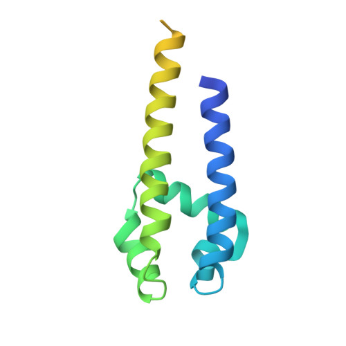
3ZJZ

Open-form NavMS Sodium Channel Pore (with C-terminal Domain)


Experimental Data Snapshot

  • Method: X-RAY DIFFRACTION
  • Resolution: 2.92 Å
  • R-Value Free: 0.287 
  • R-Value Work: 0.257 
  • R-Value Observed: 0.259 

wwPDB Validation   3D Report Full Report


Ligand Structure Quality Assessment 


This is version 1.1 of the entry. See complete history


Literature

Role of the C-Terminal Domain in the Structure and Function of Tetrameric Sodium Channels.

Bagneris, C.Decaen, P.G.Hall, B.A.Naylor, C.E.Clapham, D.E.Kay, C.W.M.Wallace, B.A.

(2013) Nat Commun 4: 2465

  • DOI: https://doi.org/10.1038/ncomms3465
  • Primary Citation of Related Structures:  
    3ZJZ

  • PubMed Abstract: 

    Voltage-gated sodium channels have essential roles in electrical signalling. Prokaryotic sodium channels are tetramers consisting of transmembrane (TM) voltage-sensing and pore domains, and a cytoplasmic carboxy-terminal domain. Previous crystal structures of bacterial sodium channels revealed the nature of their TM domains but not their C-terminal domains (CTDs). Here, using electron paramagnetic resonance (EPR) spectroscopy combined with molecular dynamics, we show that the CTD of the NavMs channel from Magnetococcus marinus includes a flexible region linking the TM domains to a four-helix coiled-coil bundle. A 2.9 Å resolution crystal structure of the NavMs pore indicates the position of the CTD, which is consistent with the EPR-derived structure. Functional analyses demonstrate that the coiled-coil domain couples inactivation with channel opening, and is enabled by negatively charged residues in the linker region. A mechanism for gating is proposed based on the structure, whereby splaying of the bottom of the pore is possible without requiring unravelling of the coiled-coil.


  • Organizational Affiliation

    1] School of Biological Sciences, Institute of Structural and Molecular Biology, Birkbeck College, University of London, London WC1E 7HX, UK [2].


Macromolecules
Find similar proteins by:  (by identity cutoff)  |  3D Structure
Entity ID: 1
MoleculeChains Sequence LengthOrganismDetailsImage
ION TRANSPORT PROTEIN
A, B, C, D
149Magnetococcus marinus MC-1Mutation(s): 0 
Membrane Entity: Yes 
UniProt
Find proteins for A0L5S6 (Magnetococcus marinus (strain ATCC BAA-1437 / JCM 17883 / MC-1))
Explore A0L5S6 
Go to UniProtKB:  A0L5S6
Entity Groups  
Sequence Clusters30% Identity50% Identity70% Identity90% Identity95% Identity100% Identity
UniProt GroupA0L5S6
Sequence Annotations
Expand
  • Reference Sequence
Small Molecules
Ligands 3 Unique
IDChains Name / Formula / InChI Key2D Diagram3D Interactions
12P
Query on 12P

Download Ideal Coordinates CCD File 
F [auth A],
K [auth B],
M [auth C]
DODECAETHYLENE GLYCOL
C24 H50 O13
WRZXKWFJEFFURH-UHFFFAOYSA-N
2CV
Query on 2CV

Download Ideal Coordinates CCD File 
E [auth A],
J [auth B],
L [auth C],
P [auth D]
HEGA-10
C18 H37 N O7
ITEIKACYSCODFV-ATLSCFEFSA-N
NA
Query on NA

Download Ideal Coordinates CCD File 
G [auth A],
H [auth A],
I [auth A],
N [auth C],
O [auth C]
SODIUM ION
Na
FKNQFGJONOIPTF-UHFFFAOYSA-N
Experimental Data & Validation

Experimental Data

  • Method: X-RAY DIFFRACTION
  • Resolution: 2.92 Å
  • R-Value Free: 0.287 
  • R-Value Work: 0.257 
  • R-Value Observed: 0.259 
  • Space Group: C 2 2 21
Unit Cell:
Length ( Å )Angle ( ˚ )
a = 80.15α = 90
b = 334.38β = 90
c = 80.21γ = 90
Software Package:
Software NamePurpose
BUSTERrefinement
XDSdata reduction
Aimlessdata scaling
PHASERphasing

Structure Validation

View Full Validation Report


Ligand Structure Quality Assessment 


Entry History 

Deposition Data

Revision History  (Full details and data files)

  • Version 1.0: 2013-10-02
    Type: Initial release
  • Version 1.1: 2023-12-20
    Changes: Data collection, Database references, Derived calculations, Other, Refinement description