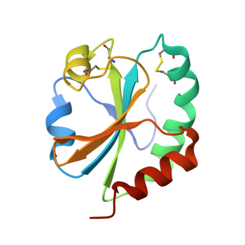
3WGE

Crystal structure of ERp46 Trx2


Experimental Data Snapshot

  • Method: X-RAY DIFFRACTION
  • Resolution: 0.95 Å
  • R-Value Free: 0.135 
  • R-Value Work: 0.122 
  • R-Value Observed: 0.123 

wwPDB Validation   3D Report Full Report


This is version 1.1 of the entry. See complete history


Literature

Radically different thioredoxin domain arrangement of ERp46, an efficient disulfide bond introducer of the mammalian PDI family

Kojima, R.Okumura, M.Masui, S.Kanemura, S.Inoue, M.Saiki, M.Yamaguchi, H.Hikima, T.Suzuki, M.Akiyama, S.Inaba, K.

(2014) Structure 22: 431-443

  • DOI: https://doi.org/10.1016/j.str.2013.12.013
  • Primary Citation of Related Structures:  
    3WGD, 3WGE, 3WGX

  • PubMed Abstract: 

    The mammalian endoplasmic reticulum (ER) contains a diverse oxidative protein folding network in which ERp46, a member of the protein disulfide isomerase (PDI) family, serves as an efficient disulfide bond introducer together with Peroxiredoxin-4 (Prx4). We revealed a radically different molecular architecture of ERp46, in which the N-terminal two thioredoxin (Trx) domains with positively charged patches near their peptide-binding site and the C-terminal Trx are linked by unusually long loops and arranged extendedly, forming an opened V-shape. Whereas PDI catalyzes native disulfide bond formation by the cooperative action of two mutually facing redox-active sites on folding intermediates bound to the central cleft, ERp46 Trx domains are separated, act independently, and engage in rapid but promiscuous disulfide bond formation during early oxidative protein folding. Thus, multiple PDI family members likely contribute to different stages of oxidative folding and work cooperatively to ensure the efficient production of multi-disulfide proteins in the ER.


  • Organizational Affiliation

    Division of Protein Chemistry, Medical Institute of Bioregulation, Kyushu University, 3-1-1 Maidashi, Higashi-ku, Fukuoka 812-8582, Japan.


Macromolecules
Find similar proteins by:  (by identity cutoff)  |  3D Structure
Entity ID: 1
MoleculeChains Sequence LengthOrganismDetailsImage
Thioredoxin domain-containing protein 5113Homo sapiensMutation(s): 0 
Gene Names: TXNDC5
EC: 5.3.4.1 (UniProt), 1.8.4 (UniProt)
UniProt & NIH Common Fund Data Resources
Find proteins for Q8NBS9 (Homo sapiens)
Explore Q8NBS9 
Go to UniProtKB:  Q8NBS9
PHAROS:  Q8NBS9
GTEx:  ENSG00000239264 
Entity Groups  
Sequence Clusters30% Identity50% Identity70% Identity90% Identity95% Identity100% Identity
UniProt GroupQ8NBS9
Sequence Annotations
Expand
  • Reference Sequence
Small Molecules
Ligands 1 Unique
IDChains Name / Formula / InChI Key2D Diagram3D Interactions
GOL
Query on GOL

Download Ideal Coordinates CCD File 
B [auth A]GLYCEROL
C3 H8 O3
PEDCQBHIVMGVHV-UHFFFAOYSA-N
Experimental Data & Validation

Experimental Data

  • Method: X-RAY DIFFRACTION
  • Resolution: 0.95 Å
  • R-Value Free: 0.135 
  • R-Value Work: 0.122 
  • R-Value Observed: 0.123 
  • Space Group: P 21 21 21
Unit Cell:
Length ( Å )Angle ( ˚ )
a = 32.58α = 90
b = 39.06β = 90
c = 87.2γ = 90
Software Package:
Software NamePurpose
HKL-2000data collection
PHASERphasing
PHENIXrefinement
MOSFLMdata reduction
SCALAdata scaling

Structure Validation

View Full Validation Report


Entry History 

Deposition Data

Revision History  (Full details and data files)

  • Version 1.0: 2014-06-25
    Type: Initial release
  • Version 1.1: 2024-11-13
    Changes: Data collection, Database references, Derived calculations, Structure summary