3VRO

Crystal structure of the tyrosine kinase binding domain of Cbl-c in complex with phospho-Src peptide


Experimental Data Snapshot

  • Method: X-RAY DIFFRACTION
  • Resolution: 1.80 Å
  • R-Value Free: 0.212 
  • R-Value Work: 0.166 
  • R-Value Observed: 0.168 

wwPDB Validation   3D Report Full Report


This is version 1.1 of the entry. See complete history


Literature

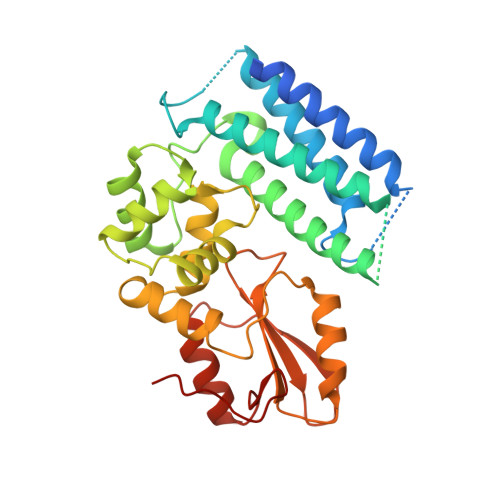
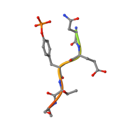
Structural flexibility regulates phosphopeptide-binding activity of the tyrosine kinase binding domain of Cbl-c.

Takeshita, K.Tezuka, T.Isozaki, Y.Yamashita, E.Suzuki, M.Kim, M.Yamanashi, Y.Yamamoto, T.Nakagawa, A.

(2012) J Biochem 152: 487-495

  • DOI: https://doi.org/10.1093/jb/mvs085
  • Primary Citation of Related Structures:  
    3VRN, 3VRO, 3VRP, 3VRQ, 3VRR

  • PubMed Abstract: 

    Through their ubiquitin ligase activity, Cbl-family proteins suppress signalling mediated by protein-tyrosine kinases (PTKs), but can also function as adaptor proteins to positively regulate signalling. The tyrosine kinase binding (TKB) domain of this family is critical for binding with tyrosine-phosphorylated target proteins. Here, we analysed the crystal structure of the TKB domain of Cbl-c/Cbl-3 (Cbl-c TKB), which is a distinct member of the mammalian Cbl-family. In comparison with Cbl TKB, Cbl-c TKB showed restricted structural flexibility upon phosphopeptide binding. A mutation in Cbl-c TKB augmenting this flexibility enhanced its binding to target phosphoproteins. These results suggest that proteins, post-translational modifications or mutations that alter structural flexibility of the TKB domain of Cbl-family proteins could regulate their binding to target phosphoproteins and thereby, affect PTK-mediated signalling.


  • Organizational Affiliation

    Institute for Protein Research, Osaka University, Suita, Osaka 565-0871, Japan.


Macromolecules
Find similar proteins by:  (by identity cutoff)  |  3D Structure
Entity ID: 1
MoleculeChains Sequence LengthOrganismDetailsImage
Signal transduction protein CBL-C331Homo sapiensMutation(s): 0 
Gene Names: CBLC
UniProt & NIH Common Fund Data Resources
Find proteins for Q9ULV8 (Homo sapiens)
Explore Q9ULV8 
Go to UniProtKB:  Q9ULV8
PHAROS:  Q9ULV8
GTEx:  ENSG00000142273 
Entity Groups  
Sequence Clusters30% Identity50% Identity70% Identity90% Identity95% Identity100% Identity
UniProt GroupQ9ULV8
Sequence Annotations
Expand
  • Reference Sequence

Find similar proteins by:  Sequence   |   3D Structure  

Entity ID: 2
MoleculeChains Sequence LengthOrganismDetailsImage
Proto-oncogene tyrosine-protein kinase Src13Homo sapiensMutation(s): 0 
EC: 2.7.10.2
UniProt & NIH Common Fund Data Resources
Find proteins for P12931 (Homo sapiens)
Explore P12931 
Go to UniProtKB:  P12931
PHAROS:  P12931
GTEx:  ENSG00000197122 
Entity Groups  
Sequence Clusters30% Identity50% Identity70% Identity90% Identity95% Identity100% Identity
UniProt GroupP12931
Sequence Annotations
Expand
  • Reference Sequence
Small Molecules
Ligands 1 Unique
IDChains Name / Formula / InChI Key2D Diagram3D Interactions
CA
Query on CA

Download Ideal Coordinates CCD File 
C [auth A]CALCIUM ION
Ca
BHPQYMZQTOCNFJ-UHFFFAOYSA-N
Modified Residues  1 Unique
IDChains TypeFormula2D DiagramParent
PTR
Query on PTR
B
L-PEPTIDE LINKINGC9 H12 N O6 PTYR
Experimental Data & Validation

Experimental Data

  • Method: X-RAY DIFFRACTION
  • Resolution: 1.80 Å
  • R-Value Free: 0.212 
  • R-Value Work: 0.166 
  • R-Value Observed: 0.168 
  • Space Group: C 2 2 21
Unit Cell:
Length ( Å )Angle ( ˚ )
a = 93.282α = 90
b = 108.493β = 90
c = 54.761γ = 90
Software Package:
Software NamePurpose
SCALEPACKdata scaling
MOLREPphasing
REFMACrefinement
PDB_EXTRACTdata extraction
DENZOdata reduction

Structure Validation

View Full Validation Report


Entry History 

Deposition Data

Revision History  (Full details and data files)

  • Version 1.0: 2013-03-06
    Type: Initial release
  • Version 1.1: 2017-11-22
    Changes: Refinement description