3VN4

Crystal structure of the exosite-containing fragment of human ADAMTS13 (P475S mutant)


Experimental Data Snapshot

  • Method: X-RAY DIFFRACTION
  • Resolution: 2.80 Å
  • R-Value Free: 0.277 
  • R-Value Work: 0.229 
  • R-Value Observed: 0.232 

Starting Model: experimental
View more details

wwPDB Validation   3D Report Full Report


Ligand Structure Quality Assessment 


This is version 3.0 of the entry. See complete history


Literature

Crystal structure and enzymatic activity of an ADAMTS-13 mutant with the East Asian-specific P475S polymorphism.

Akiyama, M.Nakayama, D.Takeda, S.Kokame, K.Takagi, J.Miyata, T.

(2013) J Thromb Haemost 11: 1399-1406

  • DOI: https://doi.org/10.1111/jth.12279
  • Primary Citation of Related Structures:  
    3VN4

  • PubMed Abstract: 

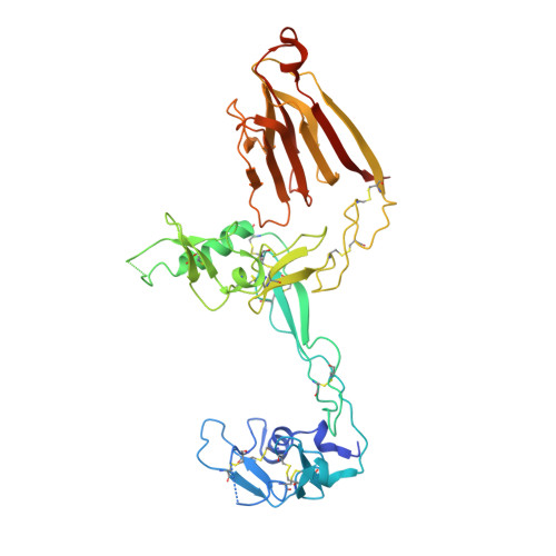
    An East Asian-specific P475S polymorphism in the gene encoding ADAMTS-13 causes an approximately 16% reduction in plasma ADAMTS-13 activity. To demonstrate the impact of this dysfunctional polymorphism by characterizing the structure and activity of the P475S mutant protein. We determined the crystal structure of the P475S mutant of ADAMTS-13-DTCS (DTCS-P475S, residues 287-685) and compared it with the wild-type structure. We determined the enzymatic parameters of ADAMTS-13-MDTCS (residues 75-685) and MDTCS-P475S, and further examined the effects of denaturants and reaction temperature on their activity. We also examined the cleavage of shear-treated von Willebrand factor (VWF) by MDTCS-P475S. MDTCS-P475S showed a reaction rate similar to that of wild-type MDTCS, but showed two-fold lower affinity for the peptidyl substrate, indicating that the Pro475-containing V-loop (residues 474-481) in the CA domain is a substrate-binding exosite. Structural analysis showed that the conformation of the V-loop was significantly different in DTCS-P475S and the wild type, where no obvious interactions of Ser475 with other residues were observed. This explains the higher susceptibility of the enzymatic activity of MDTCS-P475S to reaction environments such as denaturants and high temperature. MDTCS-P475S can moderately cleave shear-treated VWF. We have provided structural evidence that the P475S polymorphism in ADAMTS-13 leads to increased local structural instability, resulting in lowered affinity for the substrate without changing the reaction rate. The moderate activity of ADAMTS-13-P475S for shear-treated VWF is sufficient to prevent thrombotic thrombocytopenic purpura (TTP) onset.


  • Organizational Affiliation

    Department of Molecular Pathogenesis, National Cerebral and Cardiovascular Center, Osaka, Japan. akiyamam@ri.ncvc.go.jp


Macromolecules
Find similar proteins by:  (by identity cutoff)  |  3D Structure
Entity ID: 1
MoleculeChains Sequence LengthOrganismDetailsImage
A disintegrin and metalloproteinase with thrombospondin motifs 13410Homo sapiensMutation(s): 1 
Gene Names: ADAMTS13
EC: 3.4.24.87
UniProt & NIH Common Fund Data Resources
Find proteins for Q76LX8 (Homo sapiens)
Explore Q76LX8 
Go to UniProtKB:  Q76LX8
PHAROS:  Q76LX8
GTEx:  ENSG00000160323 
Entity Groups  
Sequence Clusters30% Identity50% Identity70% Identity90% Identity95% Identity100% Identity
UniProt GroupQ76LX8
Glycosylation
Glycosylation Sites: 4Go to GlyGen: Q76LX8-1
Sequence Annotations
Expand
  • Reference Sequence
Oligosaccharides

Help

Entity ID: 2
MoleculeChains Length2D Diagram Glycosylation3D Interactions
2-acetamido-2-deoxy-beta-D-glucopyranose-(1-4)-2-acetamido-2-deoxy-beta-D-glucopyranose
B
2N-Glycosylation
Glycosylation Resources
GlyTouCan:  G42666HT
GlyCosmos:  G42666HT
GlyGen:  G42666HT
Entity ID: 3
MoleculeChains Length2D Diagram Glycosylation3D Interactions
beta-D-glucopyranose-(1-3)-alpha-L-fucopyranose
C
2O-Glycosylation
Glycosylation Resources
GlyTouCan:  G36855WW
GlyCosmos:  G36855WW
GlyGen:  G36855WW
Small Molecules
Ligands 2 Unique
IDChains Name / Formula / InChI Key2D Diagram3D Interactions
NAG
Query on NAG

Download Ideal Coordinates CCD File 
D [auth A]2-acetamido-2-deoxy-beta-D-glucopyranose
C8 H15 N O6
OVRNDRQMDRJTHS-FMDGEEDCSA-N
MAN
Query on MAN

Download Ideal Coordinates CCD File 
E [auth A]alpha-D-mannopyranose
C6 H12 O6
WQZGKKKJIJFFOK-PQMKYFCFSA-N
Experimental Data & Validation

Experimental Data

  • Method: X-RAY DIFFRACTION
  • Resolution: 2.80 Å
  • R-Value Free: 0.277 
  • R-Value Work: 0.229 
  • R-Value Observed: 0.232 
  • Space Group: C 1 2 1
Unit Cell:
Length ( Å )Angle ( ˚ )
a = 153.378α = 90
b = 53.243β = 111.56
c = 76.615γ = 90
Software Package:
Software NamePurpose
HKL-2000data collection
MOLREPphasing
REFMACrefinement
HKL-2000data reduction
HKL-2000data scaling

Structure Validation

View Full Validation Report


Ligand Structure Quality Assessment 


Entry History 

Deposition Data

Revision History  (Full details and data files)

  • Version 1.0: 2012-12-26
    Type: Initial release
  • Version 2.0: 2020-07-29
    Type: Remediation
    Reason: Carbohydrate remediation
    Changes: Advisory, Atomic model, Data collection, Database references, Derived calculations, Structure summary
  • Version 2.1: 2023-11-08
    Changes: Data collection, Database references, Refinement description, Structure summary
  • Version 3.0: 2024-07-31
    Changes: Advisory, Atomic model, Data collection, Database references, Derived calculations, Non-polymer description, Structure summary