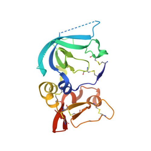
3V96

Complex of matrix metalloproteinase-10 catalytic domain (MMP-10cd) with tissue inhibitor of metalloproteinases-1 (TIMP-1)


Experimental Data Snapshot

  • Method: X-RAY DIFFRACTION
  • Resolution: 1.90 Å
  • R-Value Free: 0.266 
  • R-Value Work: 0.215 
  • R-Value Observed: 0.218 

wwPDB Validation   3D Report Full Report


This is version 1.1 of the entry. See complete history


Literature

Matrix metalloproteinase-10 (MMP-10) interaction with tissue inhibitors of metalloproteinases TIMP-1 and TIMP-2: binding studies and crystal structure.

Batra, J.Robinson, J.Soares, A.S.Fields, A.P.Radisky, D.C.Radisky, E.S.

(2012) J Biol Chem 287: 15935-15946

  • DOI: https://doi.org/10.1074/jbc.M112.341156
  • Primary Citation of Related Structures:  
    3V96

  • PubMed Abstract: 

    Matrix metalloproteinase 10 (MMP-10, stromelysin-2) is a secreted metalloproteinase with functions in skeletal development, wound healing, and vascular remodeling; its overexpression is also implicated in lung tumorigenesis and tumor progression. To understand the regulation of MMP-10 by tissue inhibitors of metalloproteinases (TIMPs), we have assessed equilibrium inhibition constants (K(i)) of putative physiological inhibitors TIMP-1 and TIMP-2 for the active catalytic domain of human MMP-10 (MMP-10cd) using multiple kinetic approaches. We find that TIMP-1 inhibits the MMP-10cd with a K(i) of 1.1 × 10(-9) M; this interaction is 10-fold weaker than the inhibition of the similar MMP-3 (stromelysin-1) catalytic domain (MMP-3cd) by TIMP-1. TIMP-2 inhibits the MMP-10cd with a K(i) of 5.8 × 10(-9) M, which is again 10-fold weaker than the inhibition of MMP-3cd by this inhibitor (K(i) = 5.5 × 10(-10) M). We solved the x-ray crystal structure of TIMP-1 bound to the MMP-10cd at 1.9 Å resolution; the structure was solved by molecular replacement and refined with an R-factor of 0.215 (R(free) = 0.266). Comparing our structure of MMP-10cd·TIMP-1 with the previously solved structure of MMP-3cd·TIMP-1 (Protein Data Bank entry 1UEA), we see substantial differences at the binding interface that provide insight into the differential binding of stromelysin family members to TIMP-1. This structural information may ultimately assist in the design of more selective TIMP-based inhibitors tailored for specificity toward individual members of the stromelysin family, with potential therapeutic applications.


  • Organizational Affiliation

    Department of Cancer Biology, Mayo Clinic Comprehensive Cancer Center, Jacksonville, Florida 32224, USA.


Macromolecules
Find similar proteins by:  (by identity cutoff)  |  3D Structure
Entity ID: 1
MoleculeChains Sequence LengthOrganismDetailsImage
Metalloproteinase inhibitor 1184Homo sapiensMutation(s): 1 
Gene Names: CLGITIMPTIMP1
UniProt & NIH Common Fund Data Resources
Find proteins for P01033 (Homo sapiens)
Explore P01033 
Go to UniProtKB:  P01033
PHAROS:  P01033
GTEx:  ENSG00000102265 
Entity Groups  
Sequence Clusters30% Identity50% Identity70% Identity90% Identity95% Identity100% Identity
UniProt GroupP01033
Sequence Annotations
Expand
  • Reference Sequence
Find similar proteins by:  (by identity cutoff)  |  3D Structure
Entity ID: 2
MoleculeChains Sequence LengthOrganismDetailsImage
Stromelysin-2165Homo sapiensMutation(s): 0 
Gene Names: MMP10STMY2
EC: 3.4.24.22
UniProt & NIH Common Fund Data Resources
Find proteins for P09238 (Homo sapiens)
Explore P09238 
Go to UniProtKB:  P09238
PHAROS:  P09238
GTEx:  ENSG00000166670 
Entity Groups  
Sequence Clusters30% Identity50% Identity70% Identity90% Identity95% Identity100% Identity
UniProt GroupP09238
Sequence Annotations
Expand
  • Reference Sequence
Small Molecules
Ligands 3 Unique
IDChains Name / Formula / InChI Key2D Diagram3D Interactions
PO4
Query on PO4

Download Ideal Coordinates CCD File 
C [auth A],
D [auth A]
PHOSPHATE ION
O4 P
NBIIXXVUZAFLBC-UHFFFAOYSA-K
ZN
Query on ZN

Download Ideal Coordinates CCD File 
E [auth B],
F [auth B]
ZINC ION
Zn
PTFCDOFLOPIGGS-UHFFFAOYSA-N
CA
Query on CA

Download Ideal Coordinates CCD File 
G [auth B],
H [auth B],
I [auth B]
CALCIUM ION
Ca
BHPQYMZQTOCNFJ-UHFFFAOYSA-N
Experimental Data & Validation

Experimental Data

  • Method: X-RAY DIFFRACTION
  • Resolution: 1.90 Å
  • R-Value Free: 0.266 
  • R-Value Work: 0.215 
  • R-Value Observed: 0.218 
  • Space Group: P 21 21 2
Unit Cell:
Length ( Å )Angle ( ˚ )
a = 231.72α = 90
b = 37.682β = 90
c = 39.523γ = 90
Software Package:
Software NamePurpose
HKL-2000data collection
PHASERphasing
REFMACrefinement
DENZOdata reduction
SCALEPACKdata scaling

Structure Validation

View Full Validation Report


Entry History 

Deposition Data

Revision History  (Full details and data files)

  • Version 1.0: 2012-03-28
    Type: Initial release
  • Version 1.1: 2012-07-25
    Changes: Database references