Visualization of Synaptic Inhibition with an Optogenetic Sensor Developed by Cell-Free Protein Engineering Automation.
Grimley, J.S., Li, L., Wang, W., Wen, L., Beese, L.S., Hellinga, H.W., Augustine, G.J.(2013) J Neurosci 33: 16297-16309
- PubMed: 24107961
- DOI: https://doi.org/10.1523/JNEUROSCI.4616-11.2013
- Primary Citation of Related Structures:
3ST0, 3SV5 - PubMed Abstract:
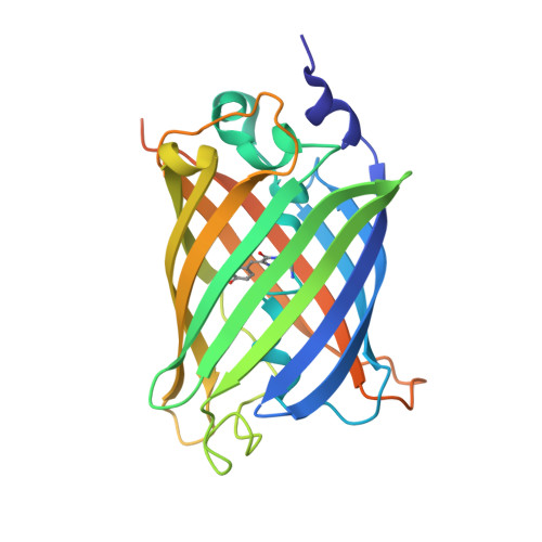
We describe an engineered fluorescent optogenetic sensor, SuperClomeleon, that robustly detects inhibitory synaptic activity in single, cultured mouse neurons by reporting intracellular chloride changes produced by exogenous GABA or inhibitory synaptic activity. Using a cell-free protein engineering automation methodology that bypasses gene cloning, we iteratively constructed, produced, and assayed hundreds of mutations in binding-site residues to identify improvements in Clomeleon, a first-generation, suboptimal sensor. Structural analysis revealed that these improvements involve halide contacts and distant side chain rearrangements. The development of optogenetic sensors that respond to neural activity enables cellular tracking of neural activity using optical, rather than electrophysiological, signals. Construction of such sensors using in vitro protein engineering establishes a powerful approach for developing new probes for brain imaging.
Organizational Affiliation:
Department of Biochemistry and Department of Neurobiology, Duke University Medical Center, Durham, North Carolina 27710, Center for Functional Connectomics, Korea Institute of Science and Technology, Seongbukgu, Seoul, 136-791 Republic of Korea, Program in Neuroscience and Behavioral Disorders, Duke-NUS Graduate Medical School, Singapore 169857, Singapore, A*STAR/Duke-NUS Neuroscience Research Partnership, Proteos, Singapore 138673, Singapore, Lee Kong Chian School of Medicine, Nanyang Technological University, Singapore 637553, Singapore, and Institute of Molecular and Cell Biology, Singapore 138673, Singapore.