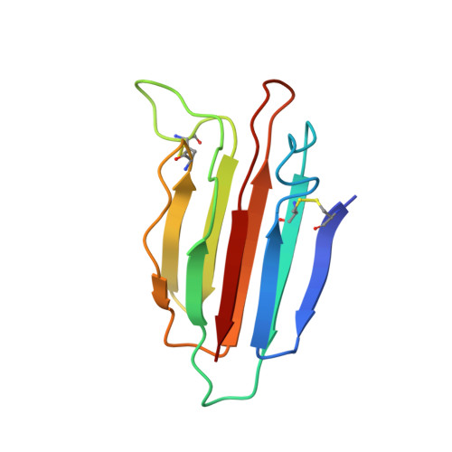
3POF

Crystal structure of MASP-1 CUB2 domain bound to Ca2+


Experimental Data Snapshot

  • Method: X-RAY DIFFRACTION
  • Resolution: 1.50 Å
  • R-Value Free: 0.149 
  • R-Value Work: 0.117 
  • R-Value Observed: 0.119 

Starting Model: experimental
View more details

wwPDB Validation   3D Report Full Report


This is version 1.3 of the entry. See complete history


Literature

Structural Basis of Mannan-Binding Lectin Recognition by Its Associated Serine Protease MASP-1: Implications for Complement Activation.

Gingras, A.R.Girija, U.V.Keeble, A.H.Panchal, R.Mitchell, D.A.Moody, P.C.Wallis, R.

(2011) Structure 19: 1635-1643

  • DOI: https://doi.org/10.1016/j.str.2011.08.014
  • Primary Citation of Related Structures:  
    3POB, 3POD, 3POE, 3POF, 3POG, 3POI, 3POJ, 3PON

  • PubMed Abstract: 

    Complement activation contributes directly to health and disease. It neutralizes pathogens and stimulates immune processes. Defects lead to immunodeficiency and autoimmune diseases, whereas inappropriate activation causes self-damage. In the lectin and classical pathways, complement is triggered upon recognition of a pathogen by an activating complex. Here we present the first structure of such a complex in the form of the collagen-like domain of mannan-binding lectin (MBL) and the binding domain of its associated protease (MASP-1/-3). The collagen binds within a groove using a pivotal lysine side chain that interacts with Ca(2+)-coordinating residues, revealing the essential role of Ca(2+). This mode of binding is prototypic for all activating complexes of the lectin and classical pathways, and suggests a general mechanism for the global changes that drive activation. The structural insights reveal a new focus for inhibitors and we have validated this concept by targeting the binding pocket of the MASP.


  • Organizational Affiliation

    Department of Biochemistry, University of Leicester, Leicester, LE1 9HN, UK.


Macromolecules
Find similar proteins by:  (by identity cutoff)  |  3D Structure
Entity ID: 1
MoleculeChains Sequence LengthOrganismDetailsImage
Mannan-binding lectin serine protease 1
A, B
115Rattus norvegicusMutation(s): 0 
Gene Names: CrarfMASP-1Masp1Masp3
EC: 3.4.21
UniProt
Find proteins for Q8CHN8 (Rattus norvegicus)
Explore Q8CHN8 
Go to UniProtKB:  Q8CHN8
Entity Groups  
Sequence Clusters30% Identity50% Identity70% Identity90% Identity95% Identity100% Identity
UniProt GroupQ8CHN8
Sequence Annotations
Expand
  • Reference Sequence
Experimental Data & Validation

Experimental Data

  • Method: X-RAY DIFFRACTION
  • Resolution: 1.50 Å
  • R-Value Free: 0.149 
  • R-Value Work: 0.117 
  • R-Value Observed: 0.119 
  • Space Group: P 21 3
Unit Cell:
Length ( Å )Angle ( ˚ )
a = 100.46α = 90
b = 100.46β = 90
c = 100.46γ = 90
Software Package:
Software NamePurpose
ADSCdata collection
PHASERphasing
REFMACrefinement
XDSdata reduction
XSCALEdata scaling

Structure Validation

View Full Validation Report


Entry History 

Deposition Data

Revision History  (Full details and data files)

  • Version 1.0: 2011-08-24
    Type: Initial release
  • Version 1.1: 2011-11-30
    Changes: Database references
  • Version 1.2: 2023-09-06
    Changes: Data collection, Database references, Derived calculations, Refinement description
  • Version 1.3: 2024-11-20
    Changes: Structure summary