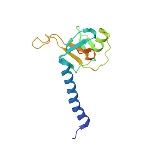
3O7X

Crystal structure of human Hili PAZ domain


Experimental Data Snapshot

  • Method: X-RAY DIFFRACTION
  • Resolution: 2.92 Å
  • R-Value Free: 0.293 
  • R-Value Work: 0.246 
  • R-Value Observed: 0.251 

wwPDB Validation   3D Report Full Report


This is version 1.3 of the entry. See complete history


Literature

Inaugural Article: Structural basis for piRNA 2'-O-methylated 3'-end recognition by Piwi PAZ (Piwi/Argonaute/Zwille) domains.

Tian, Y.Simanshu, D.K.Ma, J.B.Patel, D.J.

(2011) Proc Natl Acad Sci U S A 108: 903-910

  • DOI: https://doi.org/10.1073/pnas.1017762108
  • Primary Citation of Related Structures:  
    3O3I, 3O6E, 3O7V, 3O7X

  • PubMed Abstract: 

    Argonaute and Piwi proteins are key players in the RNA silencing pathway, with the former interacting with micro-RNAs (miRNAs) and siRNAs, whereas the latter targets piwi-interacting RNAs (piRNAs) that are 2'-O-methylated (2(')-OCH(3)) at their 3' ends. Germline-specific piRNAs and Piwi proteins play a critical role in genome defense against transposable elements, thereby protecting the genome against transposon-induced defects in gametogenesis and fertility. Humans contain four Piwi family proteins designated Hiwi1, Hiwi2, Hiwi3, and Hili. We report on the structures of Hili-PAZ (Piwi/Argonaute/Zwille) domain in the free state and Hiwi1 PAZ domain bound to self-complementary 14-mer RNAs (12-bp + 2-nt overhang) containing 2(')-OCH(3) and 2'-OH at their 3' ends. These structures explain the molecular basis underlying accommodation of the 2(')-OCH(3) group within a preformed Hiwi1 PAZ domain binding pocket, whose hydrophobic characteristics account for the preferential binding of 2(')-OCH(3) over 2'-OH 3' ends. These results contrast with the more restricted binding pocket for the human Ago1 PAZ domain, which exhibits a reverse order, with preferential binding of 2'-OH over 2(')-OCH(3) 3' ends.


  • Organizational Affiliation

    Structural Biology Program, Memorial Sloan-Kettering Cancer Center, New York, NY 10065, USA.


Macromolecules
Find similar proteins by:  (by identity cutoff)  |  3D Structure
Entity ID: 1
MoleculeChains Sequence LengthOrganismDetailsImage
Piwi-like protein 2
A, B, C, D
140Homo sapiensMutation(s): 0 
Gene Names: PIWIL2HILI
EC: 3.1.26
UniProt & NIH Common Fund Data Resources
Find proteins for Q8TC59 (Homo sapiens)
Explore Q8TC59 
Go to UniProtKB:  Q8TC59
PHAROS:  Q8TC59
GTEx:  ENSG00000197181 
Entity Groups  
Sequence Clusters30% Identity50% Identity70% Identity90% Identity95% Identity100% Identity
UniProt GroupQ8TC59
Sequence Annotations
Expand
  • Reference Sequence
Experimental Data & Validation

Experimental Data

  • Method: X-RAY DIFFRACTION
  • Resolution: 2.92 Å
  • R-Value Free: 0.293 
  • R-Value Work: 0.246 
  • R-Value Observed: 0.251 
  • Space Group: C 2 2 21
Unit Cell:
Length ( Å )Angle ( ˚ )
a = 40.535α = 90
b = 142.949β = 90
c = 226.51γ = 90
Software Package:
Software NamePurpose
DENZOdata reduction
SCALEPACKdata scaling
SHARPphasing
SOLOMONphasing
PHENIXrefinement
PDB_EXTRACTdata extraction
ADSCdata collection
HKL-2000data reduction
HKL-2000data scaling

Structure Validation

View Full Validation Report


Entry History 

Deposition Data

Revision History  (Full details and data files)

  • Version 1.0: 2011-01-12
    Type: Initial release
  • Version 1.1: 2011-07-13
    Changes: Version format compliance
  • Version 1.2: 2017-11-08
    Changes: Refinement description
  • Version 1.3: 2024-02-21
    Changes: Data collection, Database references