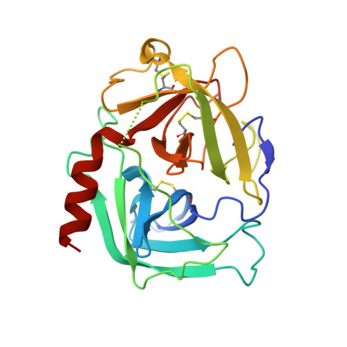
3N7O

X-ray structure of human chymase in complex with small molecule inhibitor.


Experimental Data Snapshot

  • Method: X-RAY DIFFRACTION
  • Resolution: 1.80 Å
  • R-Value Free: 0.210 
  • R-Value Work: 0.183 
  • R-Value Observed: 0.185 

Starting Model: experimental
View more details

wwPDB Validation   3D Report Full Report


Ligand Structure Quality Assessment 


This is version 2.2 of the entry. See complete history


Literature

Potency variation of small-molecule chymase inhibitors across species.

Kervinen, J.Crysler, C.Bayoumy, S.Abad, M.C.Spurlino, J.Deckman, I.Greco, M.N.Maryanoff, B.E.de Garavilla, L.

(2010) Biochem Pharmacol 80: 1033-1041

  • DOI: https://doi.org/10.1016/j.bcp.2010.06.014
  • Primary Citation of Related Structures:  
    3N7O

  • PubMed Abstract: 

    Chymases (EC 3.4.21.39) are mast cell serine proteinases that are variably expressed in different species and, in most cases, display either chymotryptic or elastolytic substrate specificity. Given that chymase inhibitors have emerged as potential therapeutic agents for treating various inflammatory, allergic, and cardiovascular disorders, it is important to understand interspecies differences of the enzymes as well as the behavior of inhibitors with them. We have expressed chymases from humans, macaques, dogs, sheep (MCP2 and MCP3), guinea pigs, and hamsters (HAM1 and HAM2) in baculovirus-infected insect cells. The enzymes were purified and characterized with kinetic constants by using chromogenic substrates. We evaluated in vitro the potency of five nonpeptide inhibitors, originally targeted against human chymase. The inhibitors exhibited remarkable cross-species variation of sensitivity, with the greatest potency observed against human and macaque chymases, with K(i) values ranging from approximately 0.4 to 72nM. Compounds were 10-300-fold less potent, and in some instances ineffective, against chymases from the other species. The X-ray structure of one of the potent phosphinate inhibitors, JNJ-18054478, complexed with human chymase was solved at 1.8A resolution to further understand the binding mode. Subtle variations in the residues in the active site that are already known to influence chymase substrate specificity can also strongly affect the compound potency. The results are discussed in the context of selecting a suitable animal model to study compounds ultimately targeted for human chymase.


  • Organizational Affiliation

    Johnson & Johnson Pharmaceutical Research and Development, Welsh and McKean Roads, Spring House, PA 19477, United States. jukkakervinen@comcast.net


Macromolecules
Find similar proteins by:  (by identity cutoff)  |  3D Structure
Entity ID: 1
MoleculeChains Sequence LengthOrganismDetailsImage
Chymase226Homo sapiensMutation(s): 2 
Gene Names: chymase (16-245)CMA1CYHCYM
EC: 3.4.21.39
UniProt & NIH Common Fund Data Resources
Find proteins for P23946 (Homo sapiens)
Explore P23946 
Go to UniProtKB:  P23946
PHAROS:  P23946
GTEx:  ENSG00000092009 
Entity Groups  
Sequence Clusters30% Identity50% Identity70% Identity90% Identity95% Identity100% Identity
UniProt GroupP23946
Glycosylation
Glycosylation Sites: 2Go to GlyGen: P23946-1
Sequence Annotations
Expand
  • Reference Sequence
Oligosaccharides

Help

Entity ID: 2
MoleculeChains Length2D Diagram Glycosylation3D Interactions
2-acetamido-2-deoxy-beta-D-glucopyranose-(1-4)-2-acetamido-2-deoxy-beta-D-glucopyranose
B, C
2N-Glycosylation
Glycosylation Resources
GlyTouCan:  G42666HT
GlyCosmos:  G42666HT
GlyGen:  G42666HT
Small Molecules
Ligands 2 Unique
IDChains Name / Formula / InChI Key2D Diagram3D Interactions
N7O
Query on N7O

Download Ideal Coordinates CCD File 
D [auth A](S)-[(1S)-1-(5-chloro-1-benzothiophen-3-yl)-2-{[(E)-2-(3,4-difluorophenyl)ethenyl]amino}-2-oxoethyl]methylphosphinic acid
C19 H15 Cl F2 N O3 P S
SEXLHAASDZPHOM-DKFQHHCZSA-N
UNL
Query on UNL

Download Ideal Coordinates CCD File 
E [auth A],
F [auth A]
Unknown ligand
SEXLHAASDZPHOM-DKFQHHCZSA-N
Binding Affinity Annotations 
IDSourceBinding Affinity
N7O PDBBind:  3N7O Ki: 72 (nM) from 1 assay(s)
BindingDB:  3N7O IC50: 58 (nM) from 1 assay(s)
Experimental Data & Validation

Experimental Data

  • Method: X-RAY DIFFRACTION
  • Resolution: 1.80 Å
  • R-Value Free: 0.210 
  • R-Value Work: 0.183 
  • R-Value Observed: 0.185 
  • Space Group: I 2 3
Unit Cell:
Length ( Å )Angle ( ˚ )
a = 124.475α = 90
b = 124.475β = 90
c = 124.475γ = 90
Software Package:
Software NamePurpose
PHENIXrefinement
CNSrefinement
HKL-2000data collection
HKL-2000data reduction
SCALEPACKdata scaling
CNSphasing

Structure Validation

View Full Validation Report


Ligand Structure Quality Assessment 


Entry History 

Deposition Data

Revision History  (Full details and data files)

  • Version 1.0: 2010-07-21
    Type: Initial release
  • Version 1.1: 2011-07-13
    Changes: Version format compliance
  • Version 1.2: 2019-07-17
    Changes: Advisory, Data collection, Database references, Derived calculations, Refinement description
  • Version 2.0: 2020-07-29
    Type: Remediation
    Reason: Carbohydrate remediation
    Changes: Atomic model, Data collection, Database references, Derived calculations, Structure summary
  • Version 2.1: 2023-09-06
    Changes: Advisory, Data collection, Database references, Refinement description, Structure summary
  • Version 2.2: 2024-10-09
    Changes: Structure summary