3KAQ

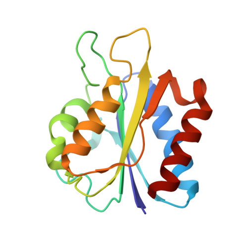
Flavodoxin from D. desulfuricans (semireduced form)


Experimental Data Snapshot

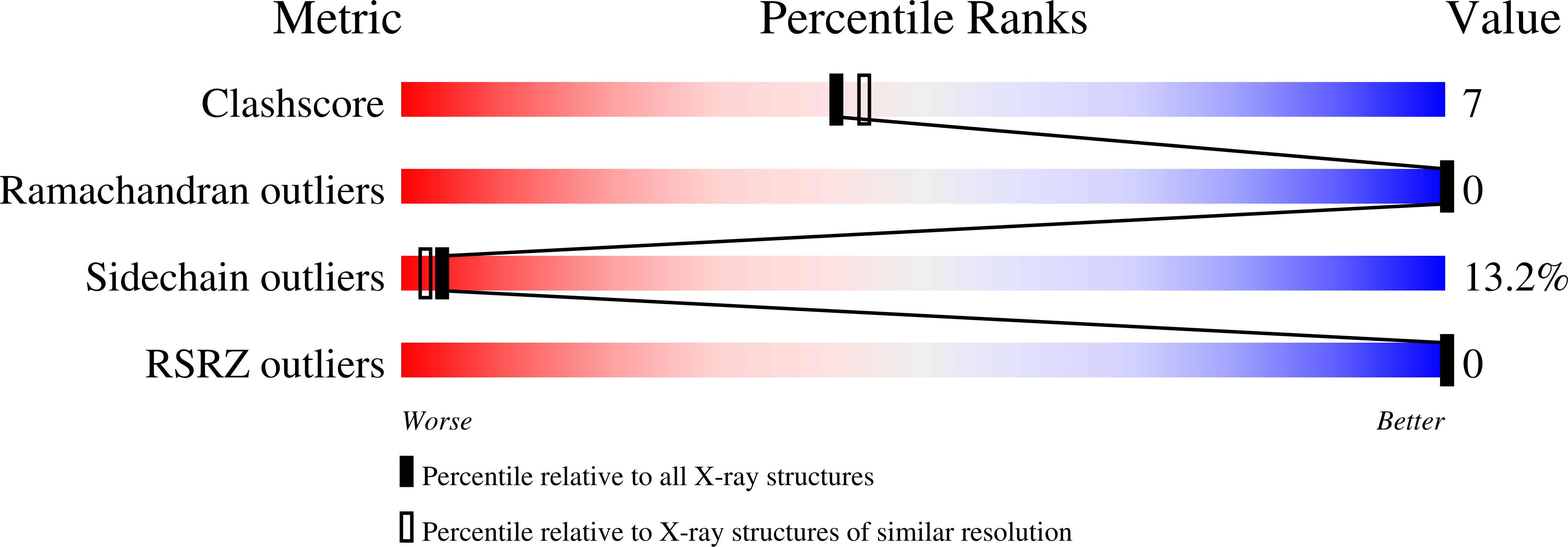
  • Method: X-RAY DIFFRACTION
  • Resolution: 2.25 Å
  • R-Value Work: 0.194 
  • R-Value Observed: 0.194 

Starting Model: experimental
View more details

wwPDB Validation   3D Report Full Report


Ligand Structure Quality Assessment 


This is version 1.4 of the entry. See complete history


Literature

Crystal structure of flavodoxin from Desulfovibrio desulfuricans ATCC 27774 in two oxidation states.

Romero, A.Caldeira, J.Legall, J.Moura, I.Moura, J.J.Romao, M.J.

(1996) Eur J Biochem 239: 190-196

  • DOI: https://doi.org/10.1111/j.1432-1033.1996.0190u.x
  • Primary Citation of Related Structures:  
    3KAP, 3KAQ

  • PubMed Abstract: 

    The crystal structures of the flavodoxin from Desulfovibrio desulfuricans ATCC 27774 have been determined and refined for both oxidized and semi-reduced forms to final crystallographic R-factors of 17.9% (0.8-0.205-nm resolution) and 19.4% (0.8-0.215-nm resolution) respectively. Native flavodoxin crystals were grown from ammonium sulfate with cell constants a = b = 9.59 nm, c=3.37nm (oxidized crystals) and they belong to space group P3(2)21. Semireduced crystals showed some changes in cell dimensions: a = b = 9.51 nm, c=3.35 nm. The three-dimensional structures are similar to other known flavodoxins and deviations are found essentially in the isoalloxazine ring environment. Conformational changes are observed between both redox states and a flip of the Gly61-Met62 peptide bond occurs upon one-electron reduction of the FMN group. These changes influence the redox potential of the oxidized/semiquinone couple. Modulation of the redox potentials is known to be related to the association constant of the FMN group to the protein. The flavodoxin from D. desulfuricans now studied has a large span between E2 (oxidized --> semiquinone) and E1 (semiquinone --> hydroquinone) redox potentials, both these values being substantially more positive within known flavodoxins. A comparison of their FMN environment was made in both oxidation states in order to correlate functional and structural differences.


  • Organizational Affiliation

    Departmento de Cristalografia, Instituto de Química Física Rocasolano, Madrid, Spain.


Macromolecules
Find similar proteins by:  (by identity cutoff)  |  3D Structure
Entity ID: 1
MoleculeChains Sequence LengthOrganismDetailsImage
Flavodoxin147Desulfovibrio desulfuricans ATCC 27774Mutation(s): 0 
UniProt
Find proteins for P80312 (Desulfovibrio desulfuricans (strain ATCC 27774 / DSM 6949 / MB))
Explore P80312 
Go to UniProtKB:  P80312
Entity Groups  
Sequence Clusters30% Identity50% Identity70% Identity90% Identity95% Identity100% Identity
UniProt GroupP80312
Sequence Annotations
Expand
  • Reference Sequence
Small Molecules
Ligands 1 Unique
IDChains Name / Formula / InChI Key2D Diagram3D Interactions
FMN
Query on FMN

Download Ideal Coordinates CCD File 
B [auth A]FLAVIN MONONUCLEOTIDE
C17 H21 N4 O9 P
FVTCRASFADXXNN-SCRDCRAPSA-N
Experimental Data & Validation

Experimental Data

  • Method: X-RAY DIFFRACTION
  • Resolution: 2.25 Å
  • R-Value Work: 0.194 
  • R-Value Observed: 0.194 
  • Space Group: P 32 2 1
Unit Cell:
Length ( Å )Angle ( ˚ )
a = 95.1α = 90
b = 95.1β = 90
c = 33.5γ = 120
Software Package:
Software NamePurpose
MAR345data collection
X-PLORmodel building
X-PLORrefinement
MOSFLMdata reduction
SCALAdata scaling
X-PLORphasing

Structure Validation

View Full Validation Report


Ligand Structure Quality Assessment 


Entry History 

Deposition Data

Revision History  (Full details and data files)

  • Version 1.0: 2009-11-24
    Type: Initial release
  • Version 1.1: 2011-07-13
    Changes: Version format compliance
  • Version 1.2: 2017-11-01
    Changes: Refinement description
  • Version 1.3: 2019-10-16
    Changes: Data collection, Database references
  • Version 1.4: 2023-11-01
    Changes: Data collection, Database references, Derived calculations, Refinement description