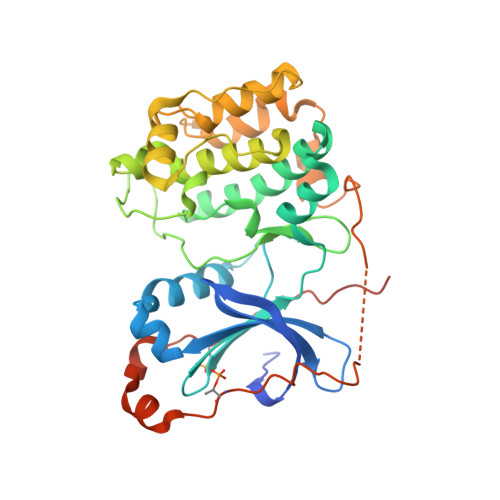
3IW4

Crystal structure of PKC alpha in complex with NVP-AEB071


Experimental Data Snapshot

  • Method: X-RAY DIFFRACTION
  • Resolution: 2.80 Å
  • R-Value Free: 0.277 
  • R-Value Work: 0.192 
  • R-Value Observed: 0.196 

wwPDB Validation   3D Report Full Report


Ligand Structure Quality Assessment 


This is version 1.3 of the entry. See complete history


Literature

Discovery of 3-(1H-indol-3-yl)-4-[2-(4-methylpiperazin-1-yl)quinazolin-4-yl]pyrrole-2,5-dione (AEB071), a potent and selective inhibitor of protein kinase C isotypes

Wagner, J.von Matt, P.Sedrani, R.Albert, R.Cooke, N.Ehrhardt, C.Geiser, M.Rummel, G.Stark, W.Strauss, A.Cowan-Jacob, S.W.Beerli, C.Weckbecker, G.Evenou, J.P.Zenke, G.Cottens, S.

(2009) J Med Chem 52: 6193-6196

  • DOI: https://doi.org/10.1021/jm901108b
  • Primary Citation of Related Structures:  
    3IW4

  • PubMed Abstract: 

    A series of novel maleimide-based inhibitors of protein kinase C (PKC) were designed, synthesized, and evaluated. AEB071 (1) was found to be a potent, selective inhibitor of classical and novel PKC isotypes. 1 is a highly efficient immunomodulator, acting via inhibition of early T cell activation. The binding mode of maleimides to PKCs, proposed by molecular modeling, was confirmed by X-ray analysis of 1 bound in the active site of PKCalpha.


  • Organizational Affiliation

    Novartis Institutes for BioMedical Research, Basel CH-4002, Switzerland. juergen.wagner@novartis.com


Macromolecules
Find similar proteins by:  (by identity cutoff)  |  3D Structure
Entity ID: 1
MoleculeChains Sequence LengthOrganismDetailsImage
Protein kinase C alpha type
A, B, C
360Homo sapiensMutation(s): 1 
Gene Names: PRKCAPKCAPRKACA
EC: 2.7.11.13
UniProt & NIH Common Fund Data Resources
Find proteins for P17252 (Homo sapiens)
Explore P17252 
Go to UniProtKB:  P17252
PHAROS:  P17252
GTEx:  ENSG00000154229 
Entity Groups  
Sequence Clusters30% Identity50% Identity70% Identity90% Identity95% Identity100% Identity
UniProt GroupP17252
Sequence Annotations
Expand
  • Reference Sequence
Small Molecules
Modified Residues  2 Unique
IDChains TypeFormula2D DiagramParent
SEP
Query on SEP
A, B, C
L-PEPTIDE LINKINGC3 H8 N O6 PSER
TPO
Query on TPO
A, B, C
L-PEPTIDE LINKINGC4 H10 N O6 PTHR
Binding Affinity Annotations 
IDSourceBinding Affinity
LW4 BindingDB:  3IW4 IC50: min: 0.95, max: 2.1 (nM) from 2 assay(s)
PDBBind:  3IW4 IC50: 2.1 (nM) from 1 assay(s)
Experimental Data & Validation

Experimental Data

  • Method: X-RAY DIFFRACTION
  • Resolution: 2.80 Å
  • R-Value Free: 0.277 
  • R-Value Work: 0.192 
  • R-Value Observed: 0.196 
  • Space Group: P 21 21 21
Unit Cell:
Length ( Å )Angle ( ˚ )
a = 43.868α = 90
b = 100.669β = 90
c = 251.335γ = 90
Software Package:
Software NamePurpose
XSCALEdata scaling
REFMACrefinement
PDB_EXTRACTdata extraction
MAR345data collection
XDSdata reduction
REFMACphasing

Structure Validation

View Full Validation Report


Ligand Structure Quality Assessment 


Entry History 

Deposition Data

Revision History  (Full details and data files)

  • Version 1.0: 2009-11-03
    Type: Initial release
  • Version 1.1: 2011-07-13
    Changes: Advisory, Refinement description, Version format compliance
  • Version 1.2: 2017-11-01
    Changes: Refinement description
  • Version 1.3: 2021-11-10
    Changes: Database references, Derived calculations