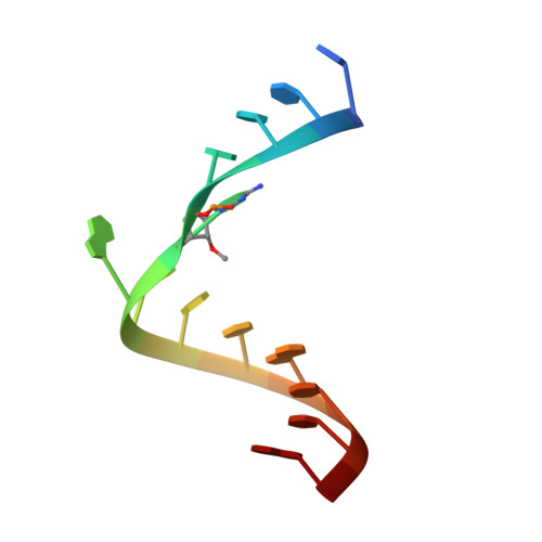
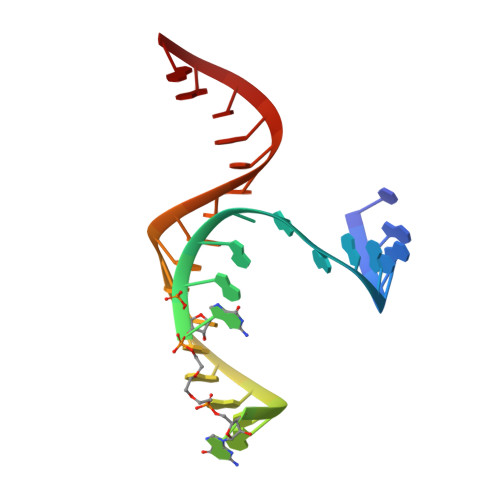
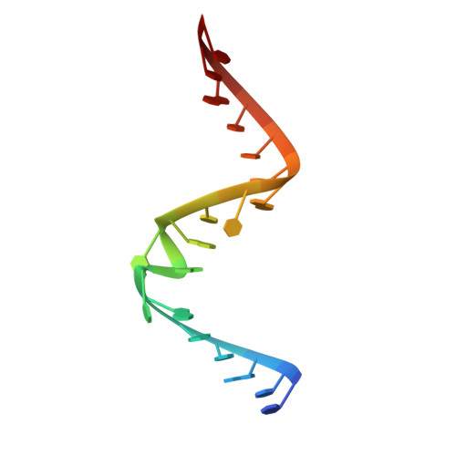
3I2S

Crystal structure of the hairpin ribozyme with a 2'OMe substrate and N1-deazaadenosine at position A10


Experimental Data Snapshot

  • Method: X-RAY DIFFRACTION
  • Resolution: 2.75 Å
  • R-Value Free: 0.261 
  • R-Value Work: 0.239 
  • R-Value Observed: 0.239 

Starting Model: experimental
View more details

wwPDB Validation   3D Report Full Report


This is version 1.2 of the entry. See complete history


Literature

Single-atom imino substitutions at A9 and A10 reveal distinct effects on the fold and function of the hairpin ribozyme catalytic core.

Spitale, R.C.Volpini, R.Mungillo, M.V.Krucinska, J.Cristalli, G.Wedekind, J.E.

(2009) Biochemistry 48: 7777-7779

  • DOI: https://doi.org/10.1021/bi9011622
  • Primary Citation of Related Structures:  
    3I2Q, 3I2R, 3I2S, 3I2U

  • PubMed Abstract: 

    The hairpin ribozyme cleaves a phosphodiester bond within a cognate substrate. Structural and biochemical data indicate the conserved A9 and A10 bases reside close to the scissile bond but make distinct contributions to catalysis. To investigate these residues, we replaced the imino moiety of each base with N1-deazaadenosine. This single-atom change resulted in an 8-fold loss in k(obs) for A9 and displacement of the base from the active site; no effects were observed for A10. We propose that the imino moiety of A9 promotes a key water-mediated contact that favors transition-state formation, which suggests an enhanced chemical repertoire for RNA.


  • Organizational Affiliation

    Department of Biochemistry & Biophysics, 601 Elmwood Avenue, Box 712, Rochester New York 14642, USA.


Macromolecules

Find similar nucleic acids by:  Sequence   |   3D Structure  

Entity ID: 1
MoleculeChains LengthOrganismImage
5'-R(*UP*CP*CP*CP*(A2M)P*GP*UP*CP*CP*AP*CP*CP*GP*U)-3'14N/A
Sequence Annotations
Expand
  • Reference Sequence
Find similar nucleic acids by: 3D Structure
Entity ID: 2
MoleculeChains LengthOrganismImage
DNA/RNA (30-MER)30N/A
Sequence Annotations
Expand
  • Reference Sequence

Find similar nucleic acids by:  Sequence   |   3D Structure  

Entity ID: 3
MoleculeChains LengthOrganismImage
5'-R(*UP*CP*GP*UP*GP*GP*UP*AP*CP*AP*UP*UP*AP*CP*CP*UP*GP*CP*C)-3'19N/A
Sequence Annotations
Expand
  • Reference Sequence
Experimental Data & Validation

Experimental Data

  • Method: X-RAY DIFFRACTION
  • Resolution: 2.75 Å
  • R-Value Free: 0.261 
  • R-Value Work: 0.239 
  • R-Value Observed: 0.239 
  • Space Group: P 61 2 2
Unit Cell:
Length ( Å )Angle ( ˚ )
a = 92.83α = 90
b = 92.83β = 90
c = 132.69γ = 120
Software Package:
Software NamePurpose
HKL-2000data collection
CNSrefinement
HKL-2000data reduction
HKL-2000data scaling
CNSphasing

Structure Validation

View Full Validation Report


Entry History 

Deposition Data

Revision History  (Full details and data files)

  • Version 1.0: 2009-11-03
    Type: Initial release
  • Version 1.1: 2011-07-13
    Changes: Non-polymer description, Version format compliance
  • Version 1.2: 2023-09-06
    Changes: Data collection, Database references, Derived calculations, Refinement description