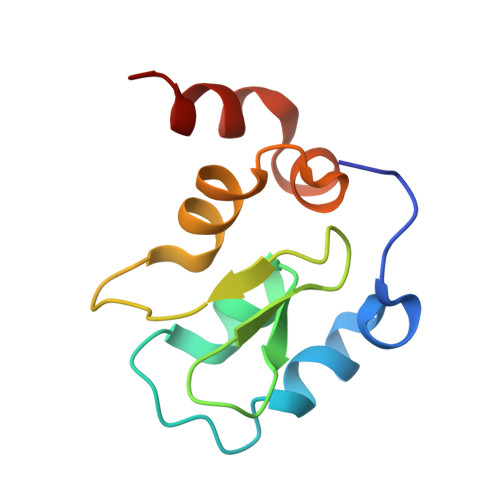
3HL5

Crystal structure of XIAP BIR3 with CS3


Experimental Data Snapshot

  • Method: X-RAY DIFFRACTION
  • Resolution: 1.80 Å
  • R-Value Free: 0.209 
  • R-Value Work: 0.176 
  • R-Value Observed: 0.179 

Starting Model: experimental
View more details

wwPDB Validation   3D Report Full Report


Ligand Structure Quality Assessment 


This is version 1.2 of the entry. See complete history


Literature

Antagonism of c-IAP and XIAP proteins is required for efficient induction of cell death by small-molecule IAP antagonists.

Ndubaku, C.Varfolomeev, E.Wang, L.Zobel, K.Lau, K.Elliott, L.O.Maurer, B.Fedorova, A.V.Dynek, J.N.Koehler, M.Hymowitz, S.G.Tsui, V.Deshayes, K.Fairbrother, W.J.Flygare, J.A.Vucic, D.

(2009) ACS Chem Biol 4: 557-566

  • DOI: https://doi.org/10.1021/cb900083m
  • Primary Citation of Related Structures:  
    3HL5

  • PubMed Abstract: 

    The inhibitor of apoptosis (IAP) proteins are critical regulators of cancer cell survival, which makes them attractive targets for therapeutic intervention in cancers. Herein, we describe the structure-based design of IAP antagonists with high affinities and selectivity (>2000-fold) for c-IAP1 over XIAP and their functional characterization as activators of apoptosis in tumor cells. Although capable of inducing cell death and preventing clonogenic survival, c-IAP-selective antagonists are significantly less potent in promoting apoptosis when compared to pan-selective compounds. However, both pan-IAP- and c-IAP-selective antagonists stimulate c-IAP1 and c-IAP2 degradation and activation of NF-kappaB pathways with comparable potencies. Therefore, although compounds that specifically target c-IAP1 and c-IAP2 are capable of inducing apoptosis, antagonism of the c-IAP proteins and XIAP is required for efficient induction of cancer cell death by IAP antagonists.


  • Organizational Affiliation

    Departments of Medicinal Chemistry and Protein Engineering, Genentech, Inc., South San Francisco, CA 94080, USA.


Macromolecules
Find similar proteins by:  (by identity cutoff)  |  3D Structure
Entity ID: 1
MoleculeChains Sequence LengthOrganismDetailsImage
Baculoviral IAP repeat-containing protein 4
A, B
95Homo sapiensMutation(s): 0 
Gene Names: API3BIRC4IAP3XIAP
EC: 6.3.2 (PDB Primary Data), 2.3.2.27 (UniProt)
UniProt & NIH Common Fund Data Resources
Find proteins for P98170 (Homo sapiens)
Explore P98170 
Go to UniProtKB:  P98170
PHAROS:  P98170
GTEx:  ENSG00000101966 
Entity Groups  
Sequence Clusters30% Identity50% Identity70% Identity90% Identity95% Identity100% Identity
UniProt GroupP98170
Sequence Annotations
Expand
  • Reference Sequence
Small Molecules
Binding Affinity Annotations 
IDSourceBinding Affinity
9JZ BindingDB:  3HL5 Ki: 3.70e+4 (nM) from 1 assay(s)
Kd: 16 (nM) from 1 assay(s)
PDBBind:  3HL5 Ki: 3.40e+4 (nM) from 1 assay(s)
Experimental Data & Validation

Experimental Data

  • Method: X-RAY DIFFRACTION
  • Resolution: 1.80 Å
  • R-Value Free: 0.209 
  • R-Value Work: 0.176 
  • R-Value Observed: 0.179 
  • Space Group: C 1 2 1
Unit Cell:
Length ( Å )Angle ( ˚ )
a = 103.913α = 90
b = 39.94β = 100
c = 49.929γ = 90
Software Package:
Software NamePurpose
BOSdata collection
PHASERphasing
REFMACrefinement
HKL-2000data reduction
HKL-2000data scaling

Structure Validation

View Full Validation Report


Ligand Structure Quality Assessment 


Entry History 

Deposition Data

Revision History  (Full details and data files)

  • Version 1.0: 2009-06-16
    Type: Initial release
  • Version 1.1: 2011-07-13
    Changes: Advisory, Refinement description, Version format compliance
  • Version 1.2: 2023-09-06
    Changes: Data collection, Database references, Derived calculations, Refinement description