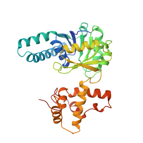
3HDH

PIG HEART SHORT CHAIN L-3-HYDROXYACYL COA DEHYDROGENASE REVISITED: SEQUENCE ANALYSIS AND CRYSTAL STRUCTURE DETERMINATION


Experimental Data Snapshot

  • Method: X-RAY DIFFRACTION
  • Resolution: 2.80 Å
  • R-Value Free: 0.287 
  • R-Value Work: 0.223 

Starting Model: experimental
View more details

wwPDB Validation   3D Report Full Report


Ligand Structure Quality Assessment 


This is version 1.3 of the entry. See complete history


Literature

Pig heart short chain L-3-hydroxyacyl-CoA dehydrogenase revisited: sequence analysis and crystal structure determination.

Barycki, J.J.O'Brien, L.K.Birktoft, J.J.Strauss, A.W.Banaszak, L.J.

(1999) Protein Sci 8: 2010-2018

  • DOI: https://doi.org/10.1110/ps.8.10.2010
  • Primary Citation of Related Structures:  
    3HDH

  • PubMed Abstract: 

    Short chain L-3-hydroxyacyl CoA dehydrogenase (SCHAD) is a soluble dimeric enzyme critical for oxidative metabolism of fatty acids. Its primary sequence has been reported to be conserved across numerous tissues and species with the notable exception of the pig heart homologue. Preliminary efforts to solve the crystal structure of the dimeric pig heart SCHAD suggested the unprecedented occurrence of three enzyme subunits within the asymmetric unit, a phenomenon that was thought to have hampered refinement of the initial chain tracing. The recently solved crystal coordinates of human heart SCHAD facilitated a molecular replacement solution to the pig heart SCHAD data. Refinement of the model, in conjunction with the nucleotide sequence for pig heart SCHAD determined in this paper, has demonstrated that the previously published pig heart SCHAD sequence was incorrect. Presented here are the corrected amino acid sequence and the high resolution crystal structure determined for pig heart SCHAD complexed with its NAD+ cofactor (2.8 A; R(cryst) = 22.4%, R(free) = 28.8%). In addition, the peculiar phenomenon of a dimeric enzyme crystallizing with three subunits contained in the asymmetric unit is described.


  • Organizational Affiliation

    Department of Biochemistry, Molecular Biology, and Biophysics, University of Minnesota, Minneapolis 55455, USA.


Macromolecules
Find similar proteins by:  (by identity cutoff)  |  3D Structure
Entity ID: 1
MoleculeChains Sequence LengthOrganismDetailsImage
PROTEIN (L-3-HYDROXYACYL COA DEHYDROGENASE)
A, B, C
302Sus scrofaMutation(s): 0 
EC: 1.1.1.35
UniProt
Find proteins for P00348 (Sus scrofa)
Explore P00348 
Go to UniProtKB:  P00348
Entity Groups  
Sequence Clusters30% Identity50% Identity70% Identity90% Identity95% Identity100% Identity
UniProt GroupP00348
Sequence Annotations
Expand
  • Reference Sequence
Experimental Data & Validation

Experimental Data

  • Method: X-RAY DIFFRACTION
  • Resolution: 2.80 Å
  • R-Value Free: 0.287 
  • R-Value Work: 0.223 
  • Space Group: C 2 2 21
Unit Cell:
Length ( Å )Angle ( ˚ )
a = 227.2α = 90
b = 82.1β = 90
c = 124.7γ = 90
Software Package:
Software NamePurpose
CNSrefinement
CNSphasing

Structure Validation

View Full Validation Report


Ligand Structure Quality Assessment 


Entry History 

Deposition Data

Revision History  (Full details and data files)

  • Version 1.0: 1999-10-08
    Type: Initial release
  • Version 1.1: 2008-04-26
    Changes: Version format compliance
  • Version 1.2: 2011-07-13
    Changes: Version format compliance
  • Version 1.3: 2023-08-02
    Changes: Database references, Derived calculations, Refinement description