3H2W

Structure of A. acidocaldarius cellulase CelA in complex with cellobiose


Experimental Data Snapshot

  • Method: X-RAY DIFFRACTION
  • Resolution: 2.66 Å
  • R-Value Free: 0.254 
  • R-Value Work: 0.193 
  • R-Value Observed: 0.196 

Starting Model: experimental
View more details

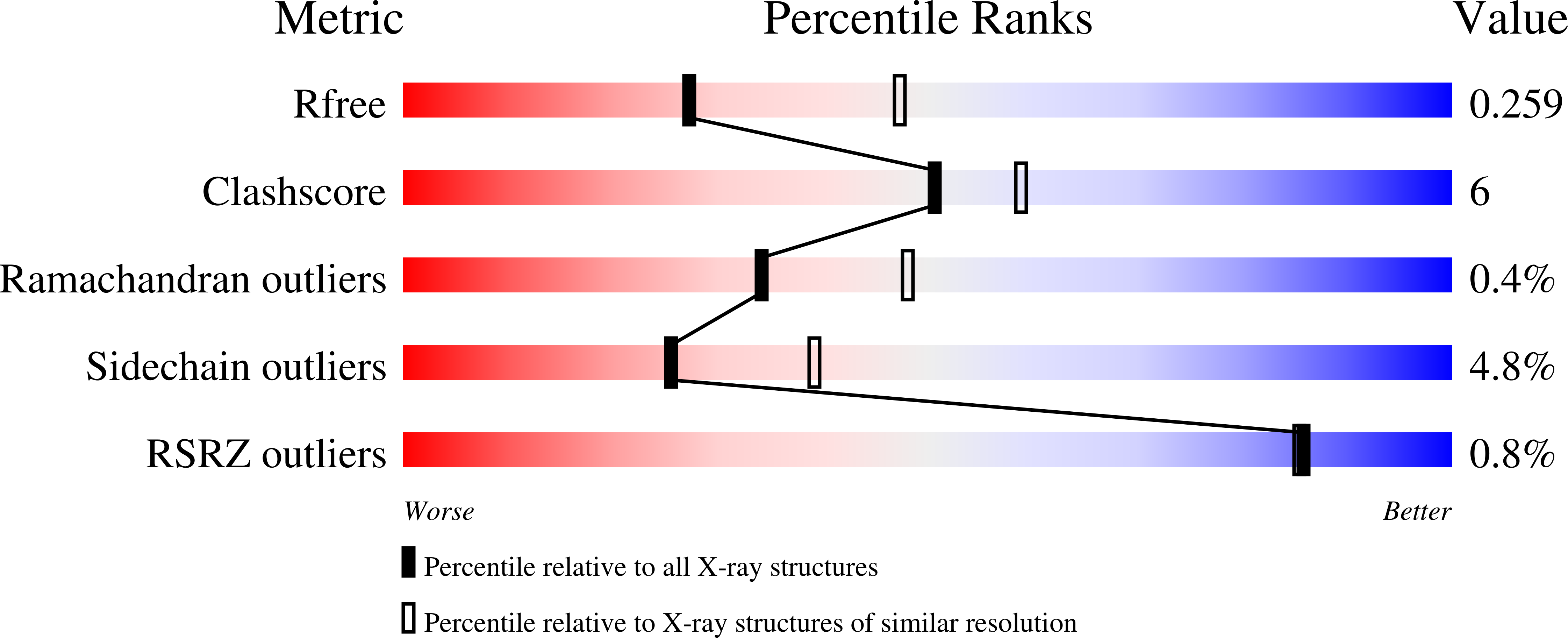
wwPDB Validation   3D Report Full Report


Ligand Structure Quality Assessment 


This is version 2.1 of the entry. See complete history


Literature

Crystal structures of A. acidocaldarius endoglucanase Cel9A in complex with Cello-oligosaccharides: strong -1 and -2 subsites mimic cellobiohydrolase activity

Eckert, K.Vigouroux, A.Lo Leggio, L.Morera, S.

(2009) J Mol Biol 394: 61-70

  • DOI: https://doi.org/10.1016/j.jmb.2009.08.060
  • Primary Citation of Related Structures:  
    3GZK, 3H2W, 3H3K

  • PubMed Abstract: 

    Alicyclobacillus acidocaldarius endoglucanase Cel9A (AaCel9A) is an inverting glycoside hydrolase with beta-1,4-glucanase activity on soluble polymeric substrates. Here, we report three X-ray structures of AaCel9A: a ligand-free structure at 1.8 A resolution and two complexes at 2.66 and 2.1 A resolution, respectively, with cellobiose obtained by co-crystallization and with cellotetraose obtained by the soaking method. AaCel9A forms an (alpha/alpha)(6)-barrel like other glycoside hydrolase family 9 enzymes. When cellobiose is used as a ligand, three glucosyl binding subsites are occupied, including two on the glycone side, while with cellotetraose as a ligand, five subsites, including four on the glycone side, are occupied. A structural comparison showed no conformational rearrangement of AaCel9A upon ligand binding. The structural analysis demonstrates that of the four minus subsites identified, subsites -1 and -2 show the strongest interaction with bound glucose. In conjunction with the open active-site cleft of AaCel9A, this is able to reconcile the previously observed cleavage of short-chain oligosaccharides with cellobiose as main product with the endo mode of action on larger polysaccharides.


  • Organizational Affiliation

    Laboratoire d'Enzymologie et Biochimie Structurales, CNRS, Avenue de la Terrasse, 91198 Gif-sur-Yvette, France.


Macromolecules
Find similar proteins by:  (by identity cutoff)  |  3D Structure
Entity ID: 1
MoleculeChains Sequence LengthOrganismDetailsImage
Cellulase537Alicyclobacillus acidocaldarius subsp. acidocaldariusMutation(s): 0 
Gene Names: celA
EC: 3.2.1.4
UniProt
Find proteins for Q9AJS0 (Alicyclobacillus acidocaldarius subsp. acidocaldarius)
Explore Q9AJS0 
Go to UniProtKB:  Q9AJS0
Entity Groups  
Sequence Clusters30% Identity50% Identity70% Identity90% Identity95% Identity100% Identity
UniProt GroupQ9AJS0
Sequence Annotations
Expand
  • Reference Sequence
Oligosaccharides

Help

Entity ID: 2
MoleculeChains Length2D Diagram Glycosylation3D Interactions
beta-D-glucopyranose-(1-4)-alpha-D-glucopyranose
B
2N/A
Glycosylation Resources
GlyTouCan:  G86802XL
GlyCosmos:  G86802XL
Biologically Interesting Molecules (External Reference) 1 Unique
Experimental Data & Validation

Experimental Data

  • Method: X-RAY DIFFRACTION
  • Resolution: 2.66 Å
  • R-Value Free: 0.254 
  • R-Value Work: 0.193 
  • R-Value Observed: 0.196 
  • Space Group: P 21 21 2
Unit Cell:
Length ( Å )Angle ( ˚ )
a = 84.892α = 90
b = 129.398β = 90
c = 48.965γ = 90
Software Package:
Software NamePurpose
JDirectordata collection
PHASERphasing
REFMACrefinement
XDSdata reduction
XDSdata scaling

Structure Validation

View Full Validation Report


Ligand Structure Quality Assessment 


Entry History 

Deposition Data

Revision History  (Full details and data files)

  • Version 1.0: 2009-10-13
    Type: Initial release
  • Version 1.1: 2011-07-13
    Changes: Version format compliance
  • Version 2.0: 2020-07-29
    Type: Remediation
    Reason: Carbohydrate remediation
    Changes: Atomic model, Data collection, Derived calculations, Structure summary
  • Version 2.1: 2023-11-01
    Changes: Data collection, Database references, Refinement description, Structure summary